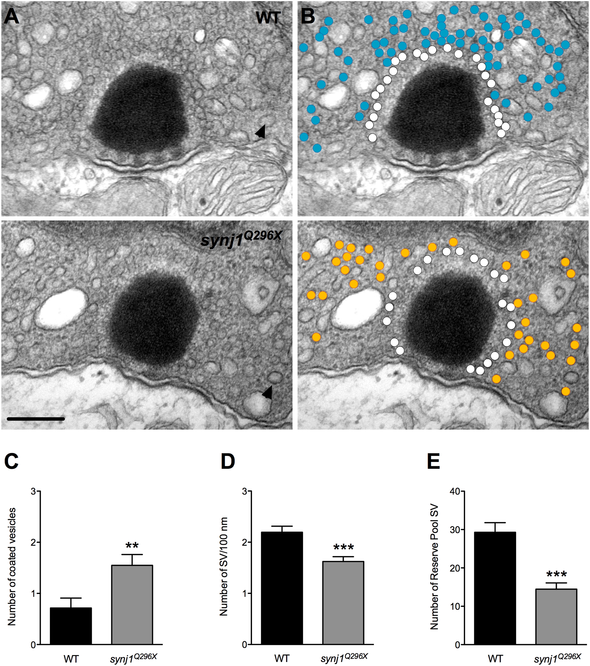
Fig. 5 Impaired vesicle recycling at ribbon synapses in mutant synj1 hair cells.
A) TEM micrographs of neuromast ribbon synapses (upper panel: wild-type ribbon; lower panel: synj1Q296X mutant ribbon). B) Same micrographs as in (A) with white circles denoting tethered vesicles. Blue and orange circles highlight the reserve vesicles in the wild type and mutant synapses, respectively. Note the reduction of reserve pool vesicles in the mutant synapse. Arrow heads indicate examples of large coated vesicles. Scale bar, 200 nm. Number of large coated vesicles (C), tethered vesicles per 100 nm of ribbon perimeter (D), and reserve pool vesicles (E) within 450 nm of ribbon centers in neuromast hair cells (black bars: wild type; grey bars: synj1Q296X).

