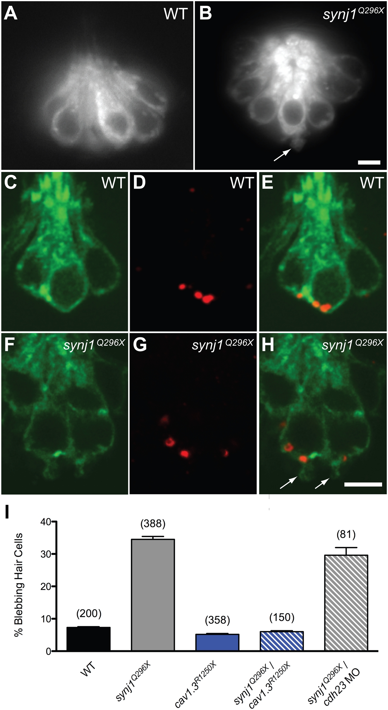
Fig. 3 Basal blebbing in synj1Q296X hair cells occurs near synaptic ribbons and is Ca2+-dependent.
FM 1–43 labeling of wild-type (A) and synj1Q296X (B) neuromast hair cells. Note the bleb or membrane protrusion at the base of a mutant cell (arrow). Single optical sections of membrane-targeted EGFP (Tg(brn3c:mgfp)) expressed in wild-type (C) and mutant (F) hair cells. D, G) Ribeye-b immunolabel (red staining) of the same neuromasts, indicating ribbons. E, H) Overlays of membrane-targeted EGFP and Ribeye-b label. Arrows in panel H indicate blebs near the ribbon in mutant hair cells. I) Basal blebbing was Ca2+-dependent. Blebbing occurred minimally in neuromast hair-cells of wild-type larvae (black bar). In contrast, blebbing was significantly increased in synj1Q296X mutant hair cells (grey bar). Blebbing was minimal in cav1.3R1250X mutant hair cells (blue bar), as well as cav1.3R1250X/synj1Q296X double mutant hair cells (blue stripe bar). Blebbing still occurred in synj1Q296X mutants that were transduction-deficient by morpholino-mediated cadherin23 knockdown (grey stripe bar). Numbers in parentheses above bars indicate number of hair cells examined. Scale bars, 3 μm.

