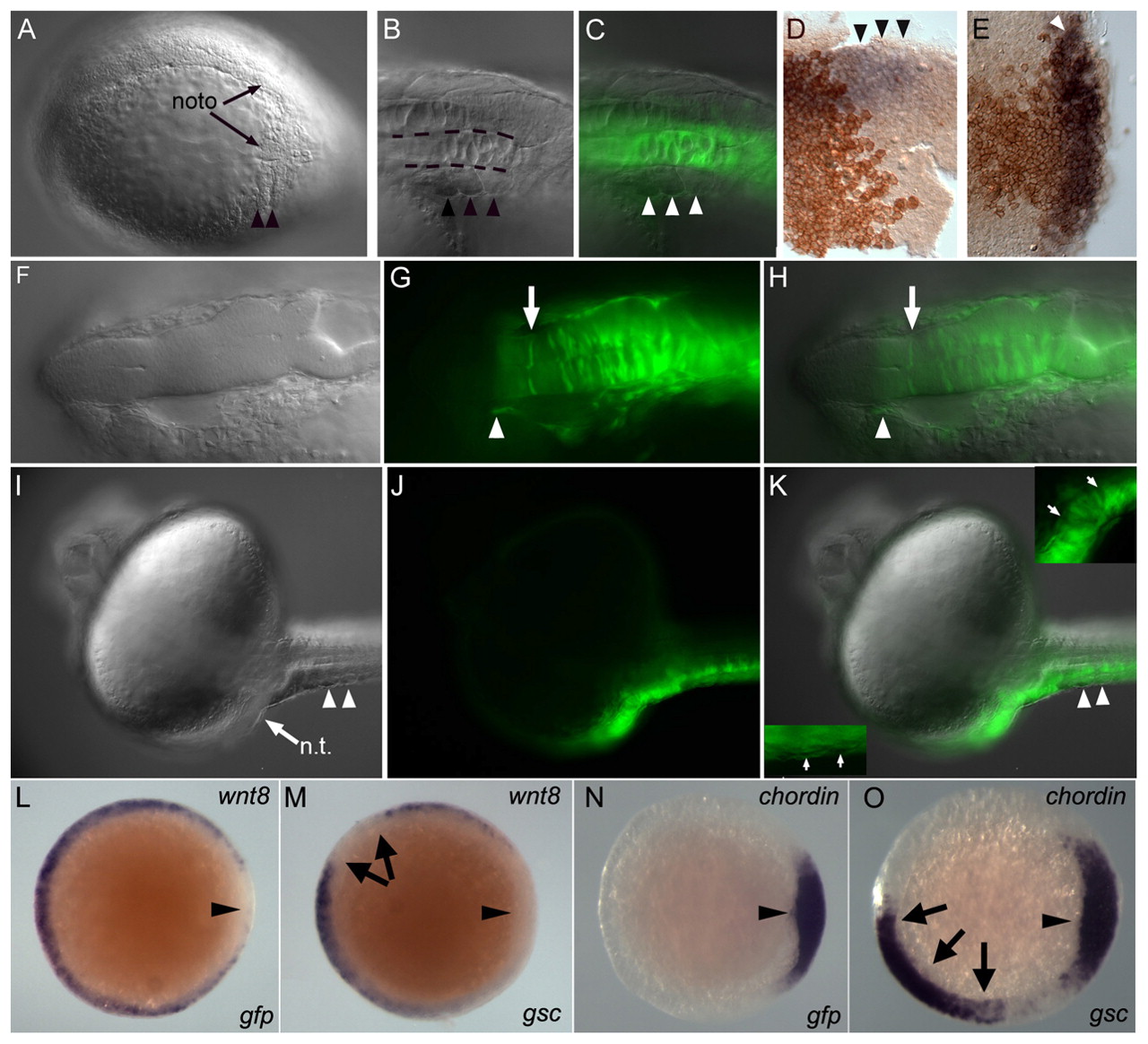
Fig. 3 gsc recruits unlabeled cells to secondary axes. (A-K) Zebrafish embryos injected with gsc RNA. (A-C) Lateral views, (A) bud; (B,C) 1 dpf. (D,E) Animal views; (F-K) dorsal views, 1 dpf. (A,B,F,I) DIC; (G,J) fluorescence; (C,H,K) merge. (A) Secondary notochord with somites (arrowheads) at bud. (B,C) Myotomes (arrowheads) with GFP-labeled notochord (dashed lines). (D,E) Shield stage gsc RNA-injected embryos stained for membrane-GFP (brown) and chd (purple), with ectopic chd (arrowheads). (F-H) Secondary neural tube with GFP-labeled (arrow) and unlabeled cells; arrowhead marks anterior GFP limit. (I-K) Low gsc RNA dose ventral clone produces partial secondary axis. (K) Arrows indicate unlabeled cells in neural tissue (right inset) and in myotomes (left inset). (L-O) Shield (arrowheads). (L,N) gfp RNA injected; (M,N) gsc RNA injected, with wnt8 (L,M) and chd (N,O) in situ probe. nt, neural tissue.
Image
Figure Caption
Acknowledgments
This image is the copyrighted work of the attributed author or publisher, and
ZFIN has permission only to display this image to its users.
Additional permissions should be obtained from the applicable author or publisher of the image.
Full text @ Development

