
Fig. 4
- ID
- ZDB-IMAGE-090429-4
- Genes
- Publication
- Yokoi et al., 2009 - Expression profiling of zebrafish sox9 mutants reveals that Sox9 is required for retinal differentiation
- All Figures
- Figures for Yokoi et al., 2009
|
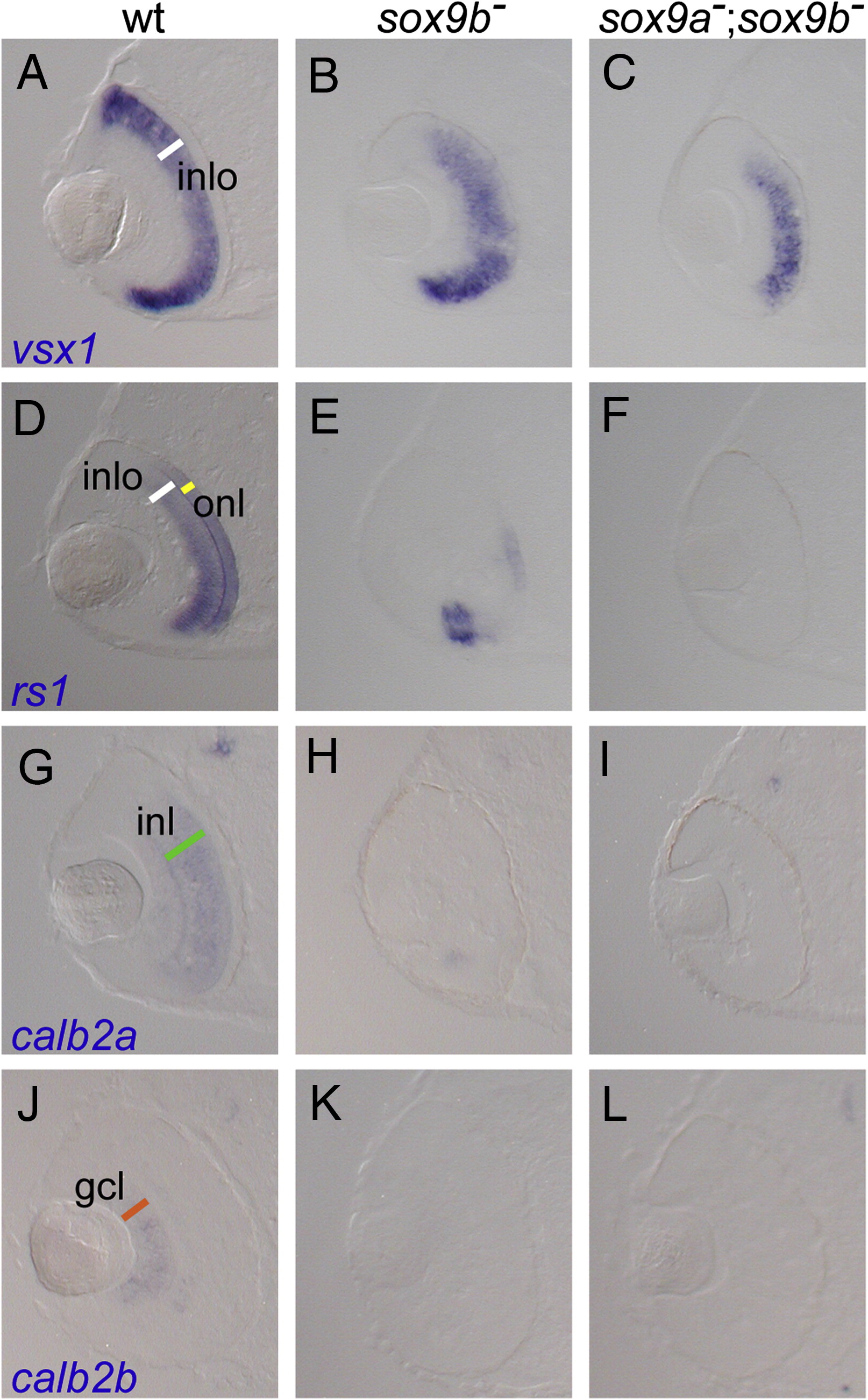
Fig. 4 Candidate genes were expressed in different layers in the developing retina. (A–C), vsx1; (D–F), rs1; (H–I), calb2a; (J–L), calb2b. Expression of vsx1 was observed in the outer part of the inner nuclear layer (A). Expression was reduced especially in the dorsal part in sox9b mutants (B) and double mutants (C). In wild-type embryos, rs1 was expressed in the outer part of the inner nuclear layer and in the outer nuclear layer (D). In sox9b mutants and sox9a;sox9b double mutant embryos, expression was severely reduced or nearly gone except for the ventral ciliary marginal zone (E, F). Expression of calb2a was observed in the inner nuclear layer (G) and calb2b was expressed in the ganglion cell layer (J). In each case, expression was severely reduced in sox9b mutants (H, K) and sox9a;sox9b double mutant embryos (I, L). White bar in (A, D), inlo, outer part of inner nuclear layer; yellow bar in (D), onl, outer nuclear layer; green bar in (G), inl, inner nuclear layer; orange bar in (J), gcl, ganglion cell layer.
Reprinted from Developmental Biology, 329(1), Yokoi, H., Yan, Y.L., Miller, M.R., Bremiller, R.A., Catchen, J.M., Johnson, E.A., and Postlethwait, J.H., Expression profiling of zebrafish sox9 mutants reveals that Sox9 is required for retinal differentiation, 1-15, Copyright (2009) with permission from Elsevier. Full text @ Dev. Biol.