
Fig. 5
- ID
- ZDB-IMAGE-090427-2
- Publication
- Paridaen et al., 2009 - Apc1-Mediated Antagonism of Wnt/beta-Catenin Signaling Is Required for Retino-Tectal Pathfinding in the Zebrafish
- All Figures
- Figures for Paridaen et al., 2009
|
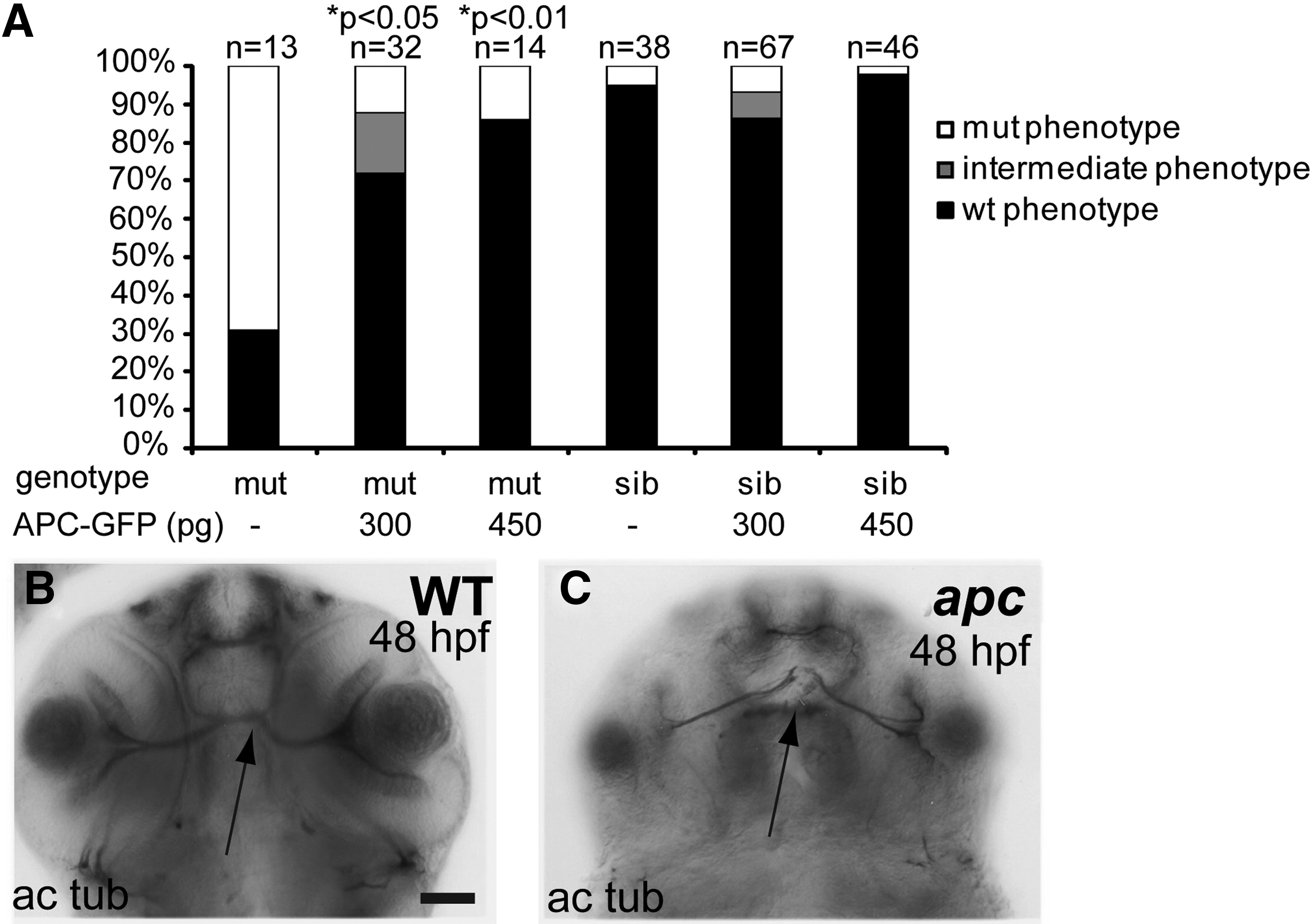
Fig. 5 Apc axon phenotype is mainly the result of β-catenin stabilization. (A) Percentage of genotyped mutant, wildtype, and heterozygous sibling embryos, showing mutant (white), intermediate (gray), or wild-type (black) phenotypes upon injection with 300 or 450 pg apc-GFP mRNA. The number of injected embryos is indicated above the bars. Injection of apc-GFP mRNA partially rescues the axonogenesis defects as demonstrated by acetylated tubulin labeling as scored over all affected brain domains. Uninjected mutant controls show variable penetrance of axon pathfinding defects (69%; n = 13). Injection with 300 pg apc-GFP causes rescue of the mutant phenotype (72%; n = 32; p<0.05). Injecting 450 pg rescues 86% of injected mutant embryos (n = 14; p<0.01). About 5% of the sibling embryos were erroneously scored as mutants. Statistics: two-tailed Fisher’s exact probability test for a table of frequency data. p-Values are indicated above the bars. (B, C) Example of a wild-type (B) and an apc mutant embryo labeled with α-acetylated tubulin antibody showing a normal and a defective ON crossing (arrow). Acetylated tubulin labeling of fully rescued apc mutants is indistinguishable from wild-type controls. (B, C) Ventral view. Scale bar = 50 μm.