
Fig. 4
- ID
- ZDB-IMAGE-090408-7
- Publication
- Cubedo et al., 2009 - CXCR4 and CXCR7 cooperate during tangential migration of facial motoneurons
- All Figures
- Figures for Cubedo et al., 2009
|
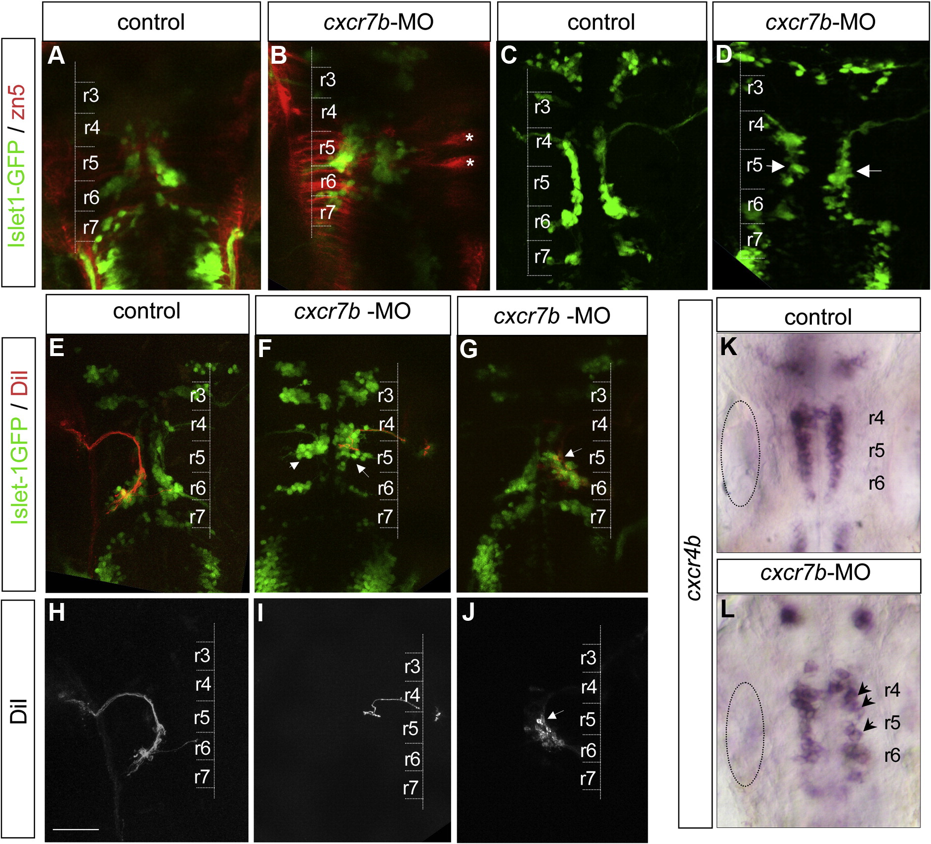
Fig. 4 cxcr7b inactivation leads to a defect of facial motoneuron migration. (A, B) zn5 immunolabeling assess GFP cell position in control (A) and cxcr7b-MO (B), zn5 is considered as a good marker for commissural neurons. r5 commissural neurons are particularly prominent (asterisks). (C, D) At 24 hpf, Islet-1-GFP expressing motoneurons form a smooth stream in control embryos but are less well organized and begin to accumulate in r5 in cxcr7b morphant (D) embryos (arrows). (E, J) Retrograde DiI labeling of facial motoneurons (red) in an Isl1-GFP background (green) at 48 hpf. The lower panels show the DiI labeling alone (H–J). (E, H) In control embryos DiI labeled cells are located mostly in r6. (F, I) In cxcr7b morphant embryos where GFP cells are positioned mainly in r5 (severe phenotype, arrows in F), DiI labeled cells are mostly found in r5. (G, J) In morphant embryos with a milder phenotype, GFP cells distributed within r5–r6 (G, arrow), DiI labeled cells are predominantly found in r5 (J). Inactivation of cxcr7b does not affect cxcr4b expression level. (K, L) The level of cxcr4b expression is similar in normal (K) and morphant (L) embryos but the pattern is disorganized in the morphants (arrows dotted circle: otic vesicle. Scale bar: 50 μm.
Reprinted from Molecular and cellular neurosciences, 40(4), Cubedo, N., Cerdan, E., Sapede, D., and Rossel, M., CXCR4 and CXCR7 cooperate during tangential migration of facial motoneurons, 474-484, Copyright (2009) with permission from Elsevier. Full text @ Mol. Cell Neurosci.