
Fig. 2
- ID
- ZDB-IMAGE-090408-5
- Publication
- Cubedo et al., 2009 - CXCR4 and CXCR7 cooperate during tangential migration of facial motoneurons
- All Figures
- Figures for Cubedo et al., 2009
|
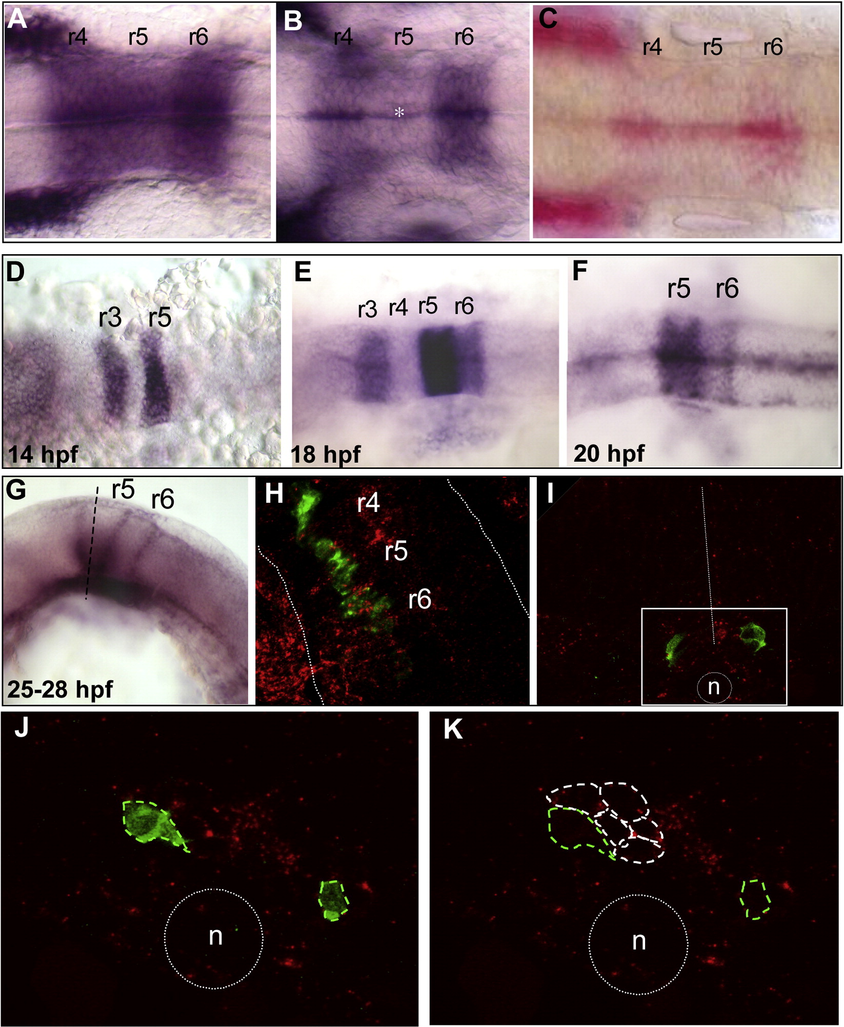
Fig. 2 sdf1a and cxcr7b expression patterns during hindbrain development. sdf1a expression (in situ hybridization) (A–C). (A) At 14 hpf, sdf1a is strongly expressed in r4, r5 and r6. (B) At 18 hpf, expression has decreased in r5 (*) and becomes almost restricted to the midline at 20 hpf (C, Fast red substrate). cxcr7b expression (D–G). (D) At 14 hpf cxcr7b is expressed in r3 and r5. (E) At 18 hpf, a strong expression is maintained in r5, but expression decreases in r3 and appears in r6. (F) By 20 hpf, expression has disappeared from r3 and decreased in r6. (G) At 24 hpf the boundaries of r5 and r6 are labeled as well as the ventral part of r5. (H) Confocal single merge image at 24 hpf (double fluorescent in situ hybridization), the detection of cxcr7b (red) and of cxcr4b (green) reveals that cxcr4b expressing cells are localized through cxcr7b expressing area (dotted white line outlined hindbrain borders). (I) Transverse section through r5 after cxcr7b in situ hybridization (red) combined with GFP immunolabeling (green). Migrating cells (GFP-positive) are localized in cxcr7b positive region (white square area), dotted white line = midline, n = notochord. (J, K) High magnification of single confocal optic section shows exclusive expressions: GFP-positive cells (green dotted line) are devoided of cxcr7b expression, which is expressed in the juxtaposed cells (white dotted line).
Reprinted from Molecular and cellular neurosciences, 40(4), Cubedo, N., Cerdan, E., Sapede, D., and Rossel, M., CXCR4 and CXCR7 cooperate during tangential migration of facial motoneurons, 474-484, Copyright (2009) with permission from Elsevier. Full text @ Mol. Cell Neurosci.