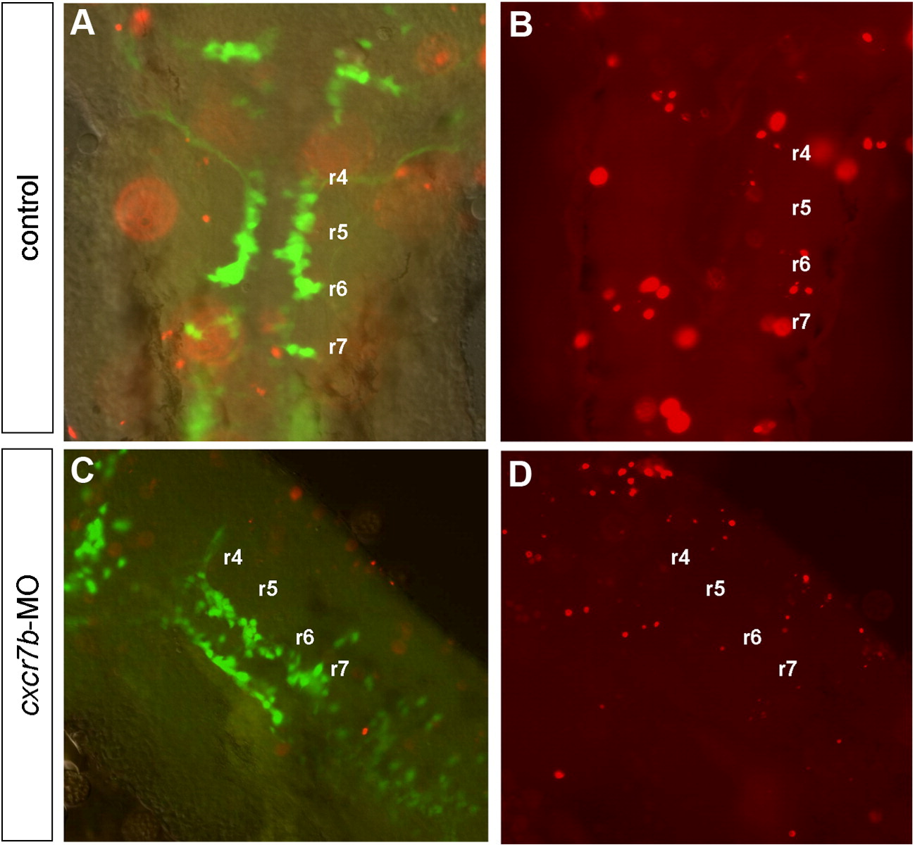
Fig. S4 Cell death analysis using the TUNEL method (red) in control (A,B) and in cxcr7b-MO (C,D) embryos at 28–30 hpf. (A,B) A few apoptotic cells are observed in control embryos in the dorsal part of the hindbrain, scattered from r2 to r7. (A,C) Merge images of GFP and TUNEL labeling at the level of facial motoneuron migration show that GFP cells have a ventral position while apoptotic cells have a dorsal position in control (A) as well as cxcr7b-MO embryo (C). (B,D) Apoptotic cells are in focus at the level of dorsal hindbrain. (C,D) No labeling was observed predominantly localized in r3 or r5 in cxcr7b-MO.
Reprinted from Molecular and cellular neurosciences, 40(4), Cubedo, N., Cerdan, E., Sapede, D., and Rossel, M., CXCR4 and CXCR7 cooperate during tangential migration of facial motoneurons, 474-484, Copyright (2009) with permission from Elsevier. Full text @ Mol. Cell Neurosci.

