
Fig. S1
- ID
- ZDB-IMAGE-090408-10
- Publication
- Cubedo et al., 2009 - CXCR4 and CXCR7 cooperate during tangential migration of facial motoneurons
- All Figures
- Figures for Cubedo et al., 2009
|
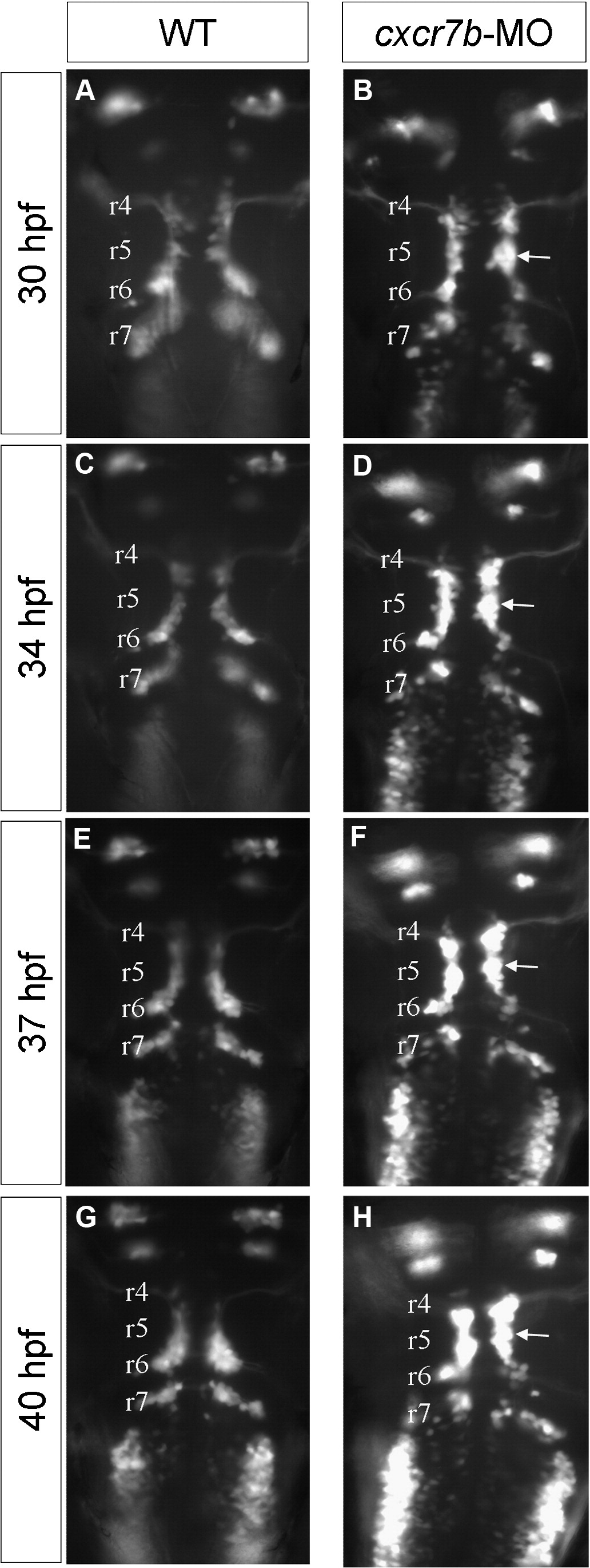
Fig. S1 Time-lapse analyses of facial motoneuron phenotype in cxcr7b-MO embryo (dorsal view). Four time points from time-lapse observations during facial motoneuron migration on Islet-1-GFP wild type (A,C,E,G) and cxcr7b morphant (B,D,F,H) embryos. The time-lapse movie extends from 28 to 42 hpf and the time-points were 30 pf (A,B), 34pf (C,D), 37 pf (E,F) and 40 pf (G,H).The total length of the hindbrain shortens during this period of time, and the correspondence between the otic vesicle and r5 is progressively lost, however the position where a large number of cells are arrested in the morphant is easily followed during the time-lapse (arrow) and can be traced back to r5.
Reprinted from Molecular and cellular neurosciences, 40(4), Cubedo, N., Cerdan, E., Sapede, D., and Rossel, M., CXCR4 and CXCR7 cooperate during tangential migration of facial motoneurons, 474-484, Copyright (2009) with permission from Elsevier. Full text @ Mol. Cell Neurosci.