
Fig. 12
- ID
- ZDB-IMAGE-090331-13
- Publication
- Nelson et al., 2009 - Retinal homeobox 1 is required for retinal neurogenesis and photoreceptor differentiation in embryonic zebrafish
- All Figures
- Figures for Nelson et al., 2009
|
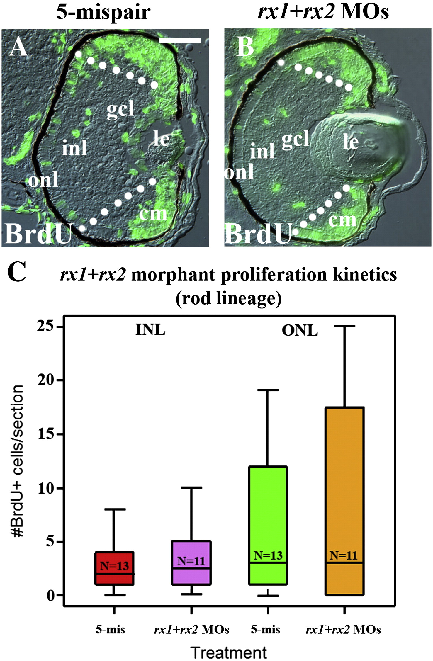
Fig. 12 Cell proliferation related to the rod lineage is unaltered following temporally-selective depletion of rx1 and rx2. (A–B) Control (A) and rx-depleted (at 44.5 hpf; B) embryos were treated with BrdU at 60 hpf, and fixed at 72 hpf for BrdU indirect immunofluorescence; patterns of BrdU incorporation are similar; BrdU-positive cells residing between the dotted lines were used for the analysis shown in C. (C) Boxplot showing numbers of BrdU-positive cells as a function of treatment and laminar position. There are no significant differences between treated and control numbers. The boxes indicate the 25th and 75th percentiles, the lines indicate the median values, and the whiskers indicate the upper and lower limits. le = lens, gcl = ganglion cell layer, inl = inner nuclear layer, onl = outer nuclear layer, cm = ciliary margin, scale bar in A = 50 μm.
Reprinted from Developmental Biology, 328(1), Nelson, S.M., Park, L., and Stenkamp, D.L., Retinal homeobox 1 is required for retinal neurogenesis and photoreceptor differentiation in embryonic zebrafish, 24-39, Copyright (2009) with permission from Elsevier. Full text @ Dev. Biol.