
Fig. S5
- ID
- ZDB-IMAGE-090224-14
- Publication
- Lam et al., 2009 - gfap and nestin reporter lines reveal characteristics of neural progenitors in the adult zebrafish brain
- All Figures
- Figures for Lam et al., 2009
|
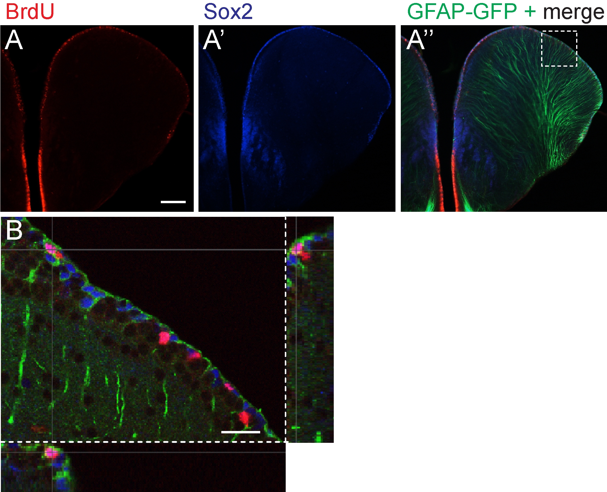
Fig. S5 The ventricular zone of the dorsal telencephalon contains mitotically active cells that express Sox 2 and glial fibrillary acidic protein (GFAP). A: Immunohistochemical staining of bromodeoxyuridine (BrdU) incorporation (red) marks mitotically active cells. A′,A″: Immunohistochemical detection of Sox2 (blue) and GFAP- green fluorescent protein (GFAP-GFP, green). A merged image (A″) shows the distribution of BrdU-labeled cells (red) in relation to GFAP and Sox 2. Images A-A″ were acquired from compound microscope. B: A confocal optical z-section, magnified from the boxed region in A″ show a proliferating cell colocalizing with Sox2 and exhibiting GFAP-positive glia processes. Dorsal up. Scale bar = 100 μm in A, 25 μm in B.