
Fig. 5
- ID
- ZDB-IMAGE-090223-30
- Genes
- Antibodies
- Publication
- Jin et al., 2009 - Definitive hematopoietic stem/progenitor cells manifest distinct differentiation output in the zebrafish VDA and PBI
- All Figures
- Figures for Jin et al., 2009
|
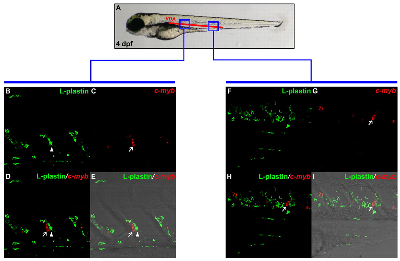
Fig. 5 HSPCs are found in the VDA in 4 dpf wild-type embryos. (A) Schematic illustration of 4 dpf embryo, indicating blue boxed regions that are magnified in B-E and F-I. (B,C) Double staining of L-plastin protein (B) and cmyb RNA (C) in anterior VDA portion of 4 dpf wild-type embryo. (D) Superimposed image of B and C. (E) Merged view of D with DIC image. (F,G) Double staining of L-plastin protein (F) and cmyb RNA (G) in posterior VDA region of 4 dpf wild-type embryo. (H) Superimposed image of F and G. (I) Merged view of H with DIC image. Arrows indicate HSPCs that are cmyb-positive only, whereas arrowheads represent L-plastin-positive myeloid cells.