
Image
|
Figure Caption
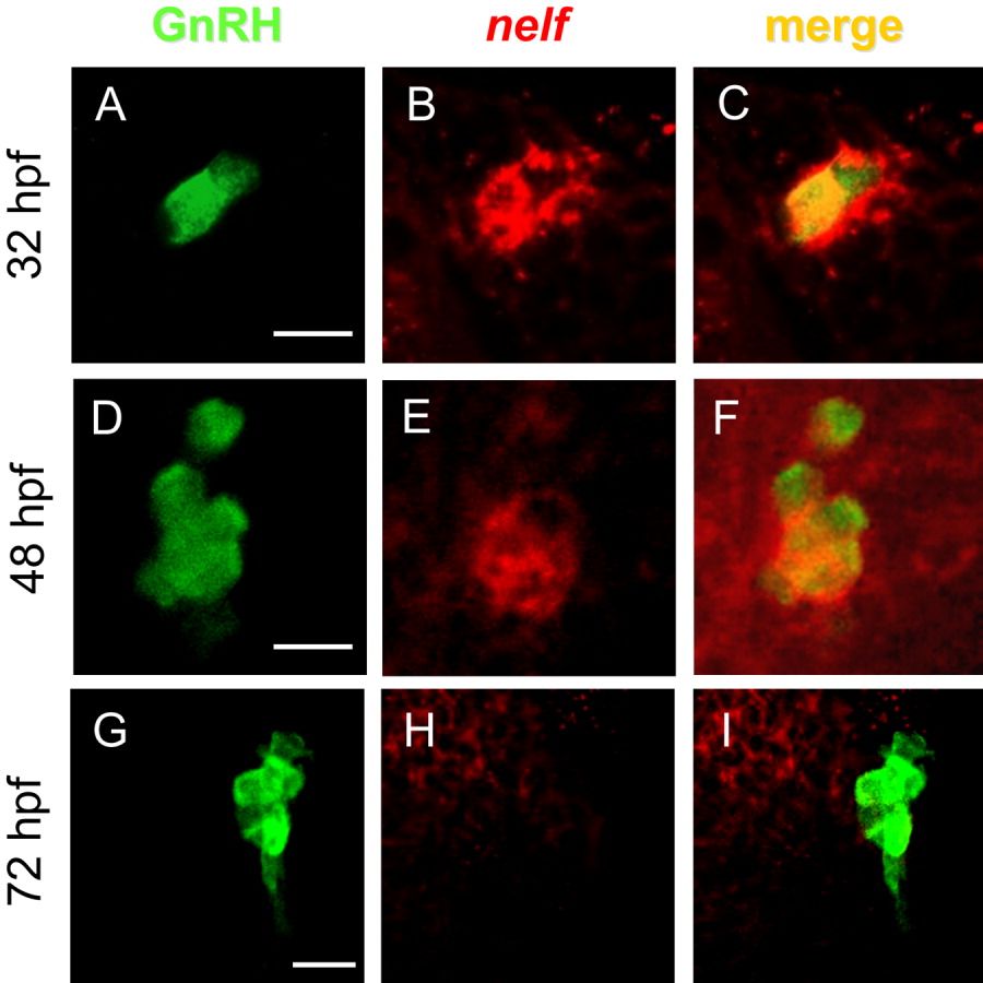
Fig. 5 GnRH3-producing neurons transiently express nelf mRNA. A-H: Staining of Tg(gnrh3:EGFP) embryos with fluorescent enhanced green fluorescent protein (EGFP) antibody (A,D,G) and nelf riboprobe (B,E,H). A single optical slice of confocal micrograph (1.8 μm). Dorsal view, anterior to the top. C,F: The analysis reveals colocalization in the olfactory epithelium at 32 hours postfertilization (hpf; C) and in the terminal nerve at 48 hpf (F). At 72 hpf, GnRH3 neurons no longer express nelf (I). Scale bar = 10 μM.
Figure Data
Acknowledgments
This image is the copyrighted work of the attributed author or publisher, and
ZFIN has permission only to display this image to its users.
Additional permissions should be obtained from the applicable author or publisher of the image.
Full text @ Dev. Dyn.