
Fig. 1
- ID
- ZDB-IMAGE-090113-17
- Genes
- Antibodies
- Publication
- Matsuda et al., 2009 - Interaction with Notch determines endocytosis of specific Delta ligands in zebrafish neural tissue
- All Figures
- Figures for Matsuda et al., 2009
|
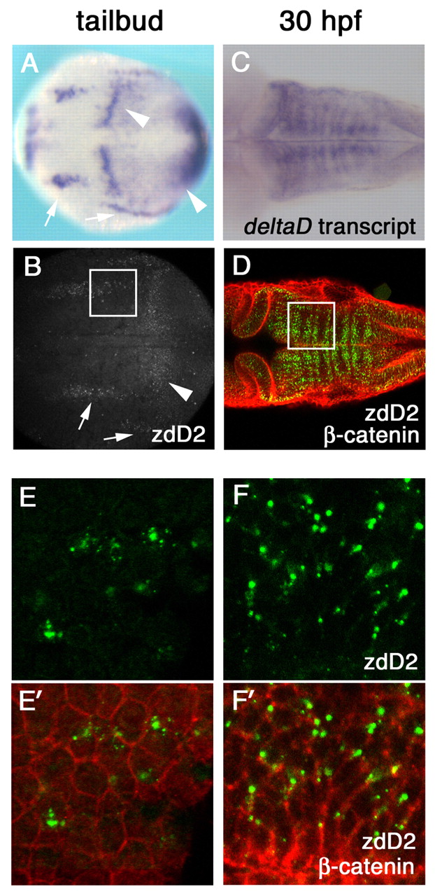
Fig. 1 DeltaD protein is primarily localized in cytoplasmic puncta. (A-D) deltaD transcript distribution (A,C) and anti-DeltaD mAb zdD2 staining (B,D) are similar. (A,B) Arrows indicate neural expression and arrowheads indicate the mesodermal expression in tail bud stage zebrafish embryos. (C,D) DeltaD expression in the hindbrain at 30 hpf. zdD2 in green and β-catenin in red. (E-F′) Magnified images correspond to squares in B and D. (E,F) zdD2 staining. (E′,F′) Merged images with zdD2 (green) and β-catenin (red). DeltaD protein is mainly detected as cytoplasmic puncta and does not colocalize with β-catenin.