
|
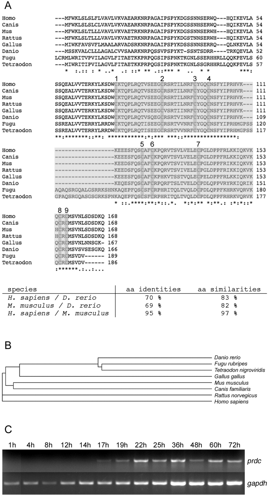
Fig. 1 The primary structure of Prdc has been highly conserved during evolution. A: Alignment of the protein related to Dan and Cerberus (PRDC) amino acid sequences from human, dog, mouse, rat, chick, zebrafish, fugu, and tetraodon. The nine conserved cysteine residues are boxed. The CAN domain is shaded. Cysteine residues 1, 3, 4, 6, 8, and 9 form the knot ring structure, 2 and 7 form a cysteine pair to complete the knot domain. Cysteine 5 is unpaired, and it might be involved in intermolecular bonds. Asterisks mark identical amino acids, double dots highly conserved substitutions, and single dots conservative amino acid changes among PRDC orthologues. The percentages of identity and similarity between human, mouse, and zebrafish are indicated below the alignment. The fugu and tetraodon sequences contain a 21-residue insertion between cysteine residues 4 and 5, which is not found in the other species. B: Phylogenetic tree of PRDC sequences from various species. C: Reverse transcriptase-polymerase chain reaction analysis using prdc gene-specific, exon-spanning primers (35 cycles). Gapdh amplification served as a control (28 cycles). The embryonic stages are indicated on top as hours postfertilization (h).