
Fig. 3
- ID
- ZDB-IMAGE-081117-20
- Genes
- Publication
- Chung et al., 2008 - Bmp2 signaling regulates the hepatic versus pancreatic fate decision
- All Figures
- Figures for Chung et al., 2008
|
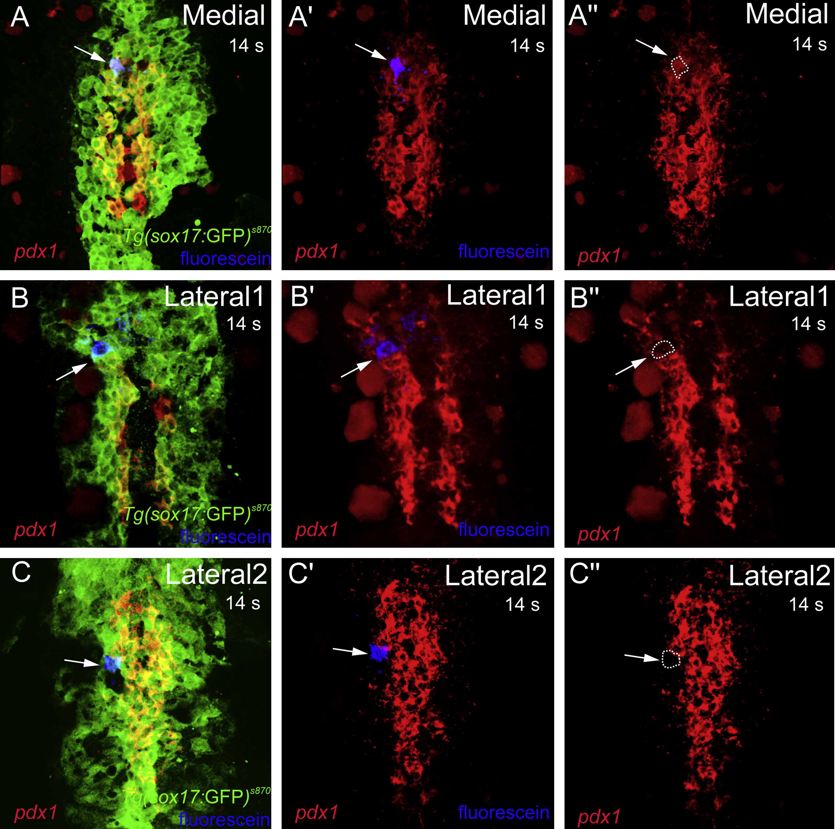
Fig. 3 The pdx1 “Gradient” Correlates with the Medio-Lateral Position of Endodermal Cells
(A–C″) Confocal projections of Tg(sox17:GFP)s870 embryos at the 14 somite stage stained for pdx1 (red), GFP (green), and uncaged-fluorescein (blue). A medial cell (A–A″) (arrow), which gives rise to the endocrine pancreas, expresses high levels of pdx1 (outlined by white dashed line in [A″]). A lateral 1 cell (B–B″) (arrow), which gives rise to the exocrine pancreas and intestine as well as a small number of pancreatic endocrine cells, expresses low levels of pdx1 (outlined by white dashed line in [B″]). A lateral 2 cell (C–C″) (arrow), which gives rise to the liver, intestine, and exocrine pancreas, does not express pdx1 (outlined by white dashed line in [C″]).
Reprinted from Developmental Cell, 15(5), Chung, W.S., Shin, C.H., and Stainier, D.Y., Bmp2 signaling regulates the hepatic versus pancreatic fate decision, 738-748, Copyright (2008) with permission from Elsevier. Full text @ Dev. Cell