
Fig. 6
- ID
- ZDB-IMAGE-081028-11
- Genes
- Publication
- Korzh et al., 2008 - Requirement of vasculogenesis and blood circulation in late stages of liver growth in zebrafish
- All Figures
- Figures for Korzh et al., 2008
|
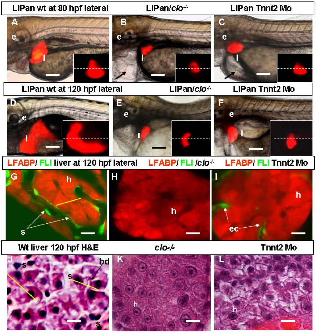
Fig. 6 Role of circulation in liver development. (A-F) Left-lateral views: liver morphogenesis in live LiPan wild type (A, D), LiPan/clo-/- mutants (B, E) and LiPan/Tnnt2 morphants (C, F) at 80 hpf (A-C) and 120 hpf (D-F). Dorsal views of the liver region of the same embryos are shown as inserts in each panel and dash lines indicate the midline with the right side at the top. Livers in clo-/- mutants and Tnnt2 morphants are located more medial, lack the anterio-ventral and posterior expansion, and are significantly reduced in size compared to the livers in wild type sibling. The cardiac edema in both mutants and morphants is indicated by arrow. (G-I) Left-lateral confocal in vivo projections of liver of LiPan/Tg(fli1:EGFP)y1 larvae at 120 hpf in wild type (G), clo-/- mutant (H) and Tnnt2 morphant (I) backgrounds. In clo-/ mutants ECs are absent (H), whereas in tnnt2 morphants they are present only between hepatocytes of the outer layer (I). (J-L) High-resolution light micrographs of hepatic parenchyma of zebrafish larvae stained with H&E. In 120-hpf wild type sibling, hepatocyte tubules are separated by sinusoids containing erythrocytes (J); in contrast, in 120-hpf clo-/- mutant (K) and Tnnt2 morphant (L), hepatocytes are tightly connected to each other. Note two sinusoids separated by two rows of neighboring hepatocytes as defined by yellow lines. Abbreviations: ec, endothelial cells; e, ear; h, hepatocytes; l, liver; s, sinusoid. In all images, anterior is towards the left. Scale bars, 125 μm in (A-F) and 625 μm in (G-L).