
Fig. 2
- ID
- ZDB-IMAGE-081003-2
- Genes
- Publication
- Luo et al., 2008 - Nr4a2 is essential for the differentiation of dopaminergic neurons during zebrafish embryogenesis
- All Figures
- Figures for Luo et al., 2008
|
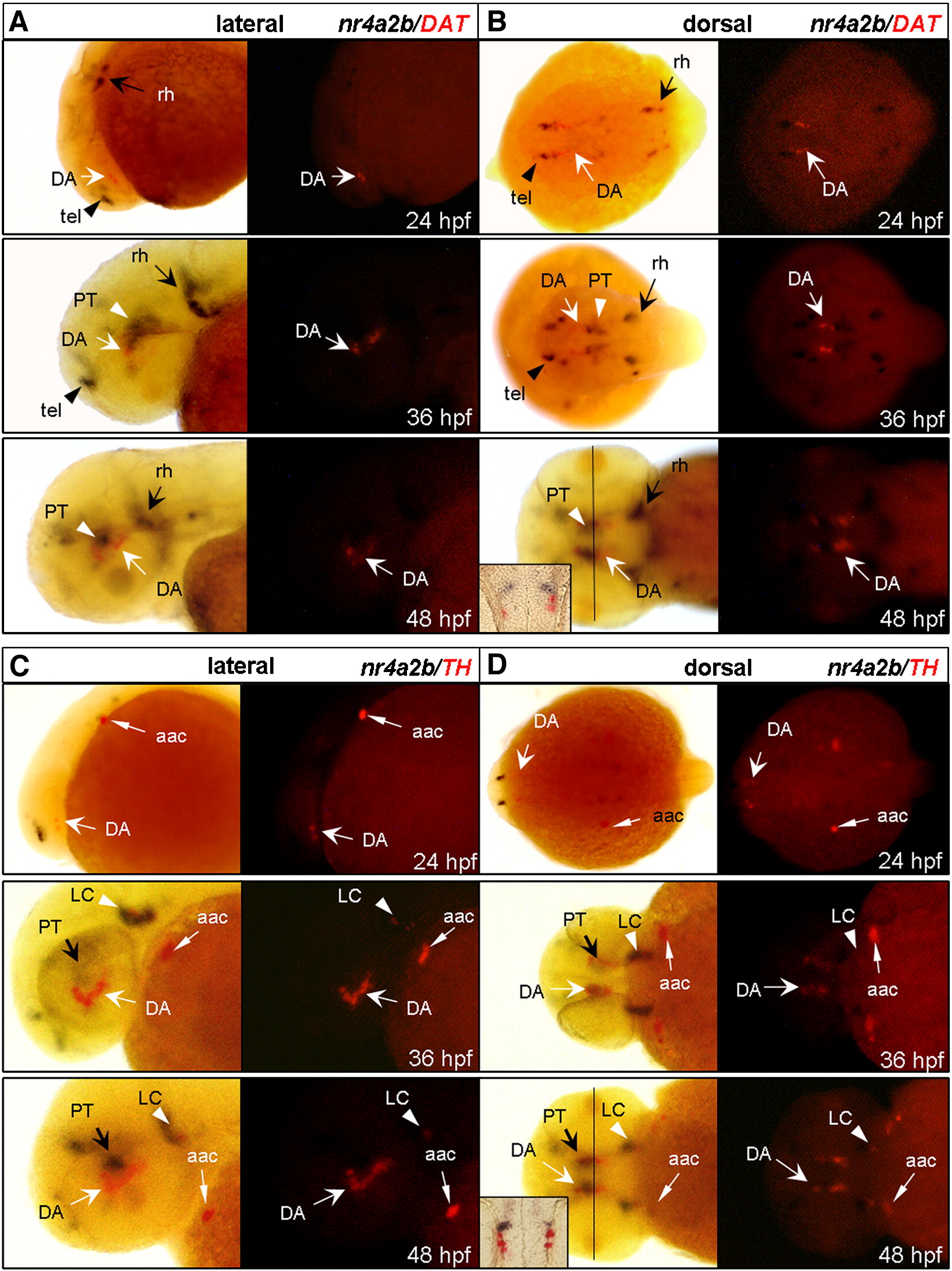
Fig. 2 nr4a2b (black) has little co-expression with TH and DAT (red) in the PT area. Anterior is to the left and dorsal is up. (A and B) Lateral and dorsal views show the nr4a2b and DAT expression patterns of 24, 36, 48 hpf embryos. DA neurons (white arrows) are located in the PT area, while nr4a2b+ cells are expressed at the dorsal (white arrowheads). (C and D) Lateral and dorsal views show the nr4a2b and TH expression pattern of 24, 36, 48 hpf embryos. Most of the TH+ neurons (white arrows) surround by nr4a2b+ cells (black arrows) in basal forebrain and some of them are closely located with nr4a2b+ cells in LC (white arrowheads). Solid lines in B and D correspond to transverse section presented as the insets. Dorsal is up. Fast Red labeling of TH+ or DAT+ cells (RITC filter set) is shown at the right panel. aac, arch-associated catecholamine cells; DA, dopamine neuron; DAT, dopamine transporter; LC, locus coeruleus; PT, posterior tuberculum; rh, rhombencephalon; tel, telencephalon; TH, tyrosine hydroxylase.
Reprinted from Molecular and cellular neurosciences, 39(2), Luo, G.R., Chen, Y., Li, X.P., Liu, T.X., and Le, W.D., Nr4a2 is essential for the differentiation of dopaminergic neurons during zebrafish embryogenesis, 202-210, Copyright (2008) with permission from Elsevier. Full text @ Mol. Cell Neurosci.