
|
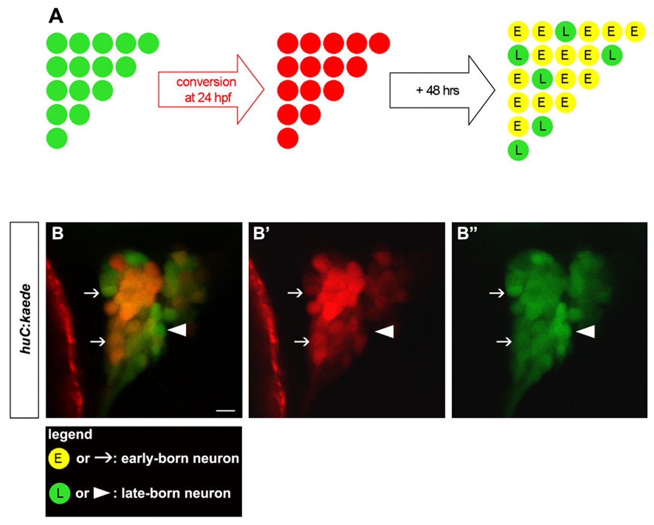
Fig. 2 In vivo birthdating analysis of trigeminal sensory neurons using BAPTI. Embryos carrying the huc:kaede transgene were analyzed using the birthdating analysis by photoconverted fluorescent protein tracing in vivo method (BAPTI). (A) As schematized, the trigeminal sensory neurons initially appear green. Following illumination of 24 hpf embryos with ultraviolet light, huc:kaedegreen is converted to huc:kaedered and all neurons born prior to 24 hpf appear red. After 48 hours incubation, neurons born before 24 hpf retain huc:kaedered and express de novo huc:kaedegreen, whereas neurons born after 24 hpf express only huc:kaedegreen. Early-born neurons (E) appear red and green (yellow); late-born (L) neurons appear green only. (B) Converted embryos were imaged at 72 hpf. Early-born neurons are identifiable by their red and green signals (arrow), whereas late-born neurons are identifiable by their green only signal (arrowhead). Neurons with weak (top arrow) or strong red signals (bottom arrow) were counted as early-born neurons. The entire trigeminal sensory ganglion was imaged by confocal microscopy. At this plane of confocal section only one late-born neuron is visible (arrowhead). Side view, anterior towards the left. Scale bar: 10 μm.