
Fig. 3
- ID
- ZDB-IMAGE-080919-6
- Genes
- Publication
- Cretekos et al., 1999 - alyron, an insertional mutation affecting early neural crest development in zebrafish
- All Figures
- Figures for Cretekos et al., 1999
|
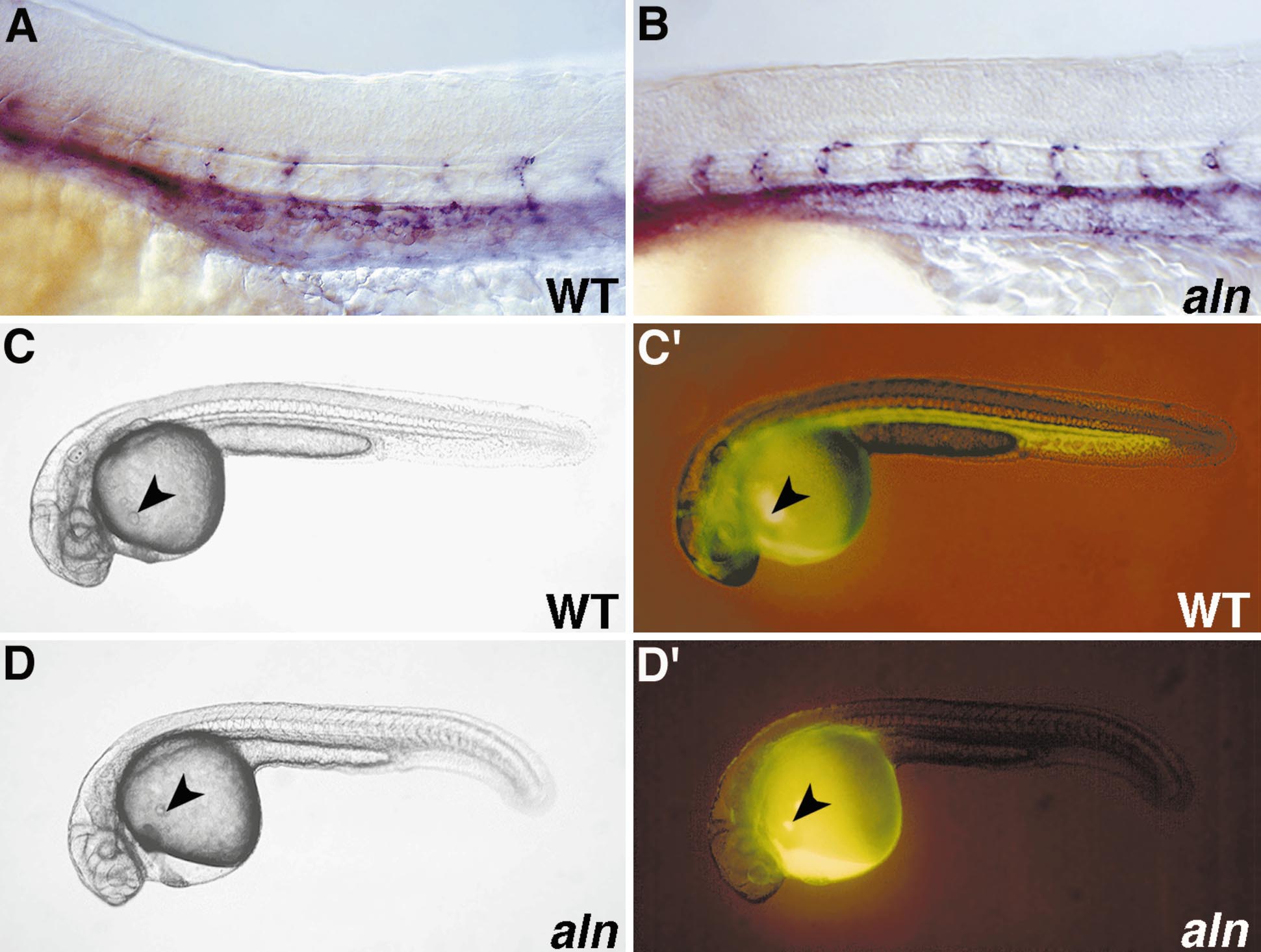
Fig. 3 Analysis of cardiovascular defect. (A, B) WT sibling (A) and aln mutant (B) 24-h embryos stained for flk-1 expression by RNA in situ hybridization. The major blood vessels appear to be formed normally in mutant embryos. (C–D′) Fluid circulation was visualized by microinjection of fluorescent dye into the common cardinal vein. The site of dye injection in a 26-h WT sibling (C, C′) and aln mutant (D, D′) is indicated by arrowheads. (C′) Epifluorescence view of the WT embryo pictured in C, photographed about 1 min after injection. Dye has passed through the heart, circulated through the head, and traveled to and returned from the tail. (D′) Epifluorescence view of the aln mutant embryo pictured in D, photographed 30 min after injection. Dye was drawn into the heart, but little if any circulated further.
Reprinted from Developmental Biology, 210(2), Cretekos, C.J. and Grunwald, D.J., alyron, an insertional mutation affecting early neural crest development in zebrafish, 322-338, Copyright (1999) with permission from Elsevier. Full text @ Dev. Biol.