
Fig. 3
- ID
- ZDB-IMAGE-080919-12
- Genes
- Publication
- Fürthauer et al., 1999 - Three different noggin genes antagonize the activity of bone morphogenetic proteins in the zebrafish embryo
- All Figures
- Figures for Fürthauer et al., 1999
|
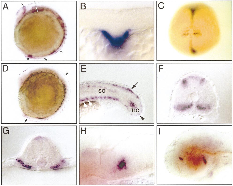
Fig. 3 Noggin1 expression at segmentation and pharyngula stages. (A) Lateral view of a 6-somite stage embryo shows nog1 expression in the posterior eye field (black arrow), the somites as well as the unsegmented paraxial mesoderm (open arrowhead), the caudal floor plate (arrowhead), and the posterior tip of the notochord (star). (B) Optical cross section of the embryo at the level indicated by the white arrow in (A). Nog1 is expressed in the hypoblast at the level of the prospective pharyngeal endoderm. (C) Dorsal view of a 3-somite-stage embryo. Note that axial nog1 expression is vanishing at the level of the forming somites, while expression is still robust at more anterior or posterior levels. (D) Lateral view of a 12-somite-stage embryo. Nog1 becomes detectable in the dorsal neural tube from the diencephalon caudalward (arrowhead). Faint expression is still observed in the caudal floor plate (arrow). (E, F) At the 18-somite stage, nog1 is expressed in discrete dorsal and ventral aspects of the recently formed caudal somites (so). In older more anterior somites only nog1 is expressed only ventrally (white arrowheads). Caudally, nog1 is expressed in the dorsal spinal cord (arrow), the tip of the notochord (nc), and the mesenchyme lining the tail bud (black arrowhead). (G) At 24 h of development, somitic nog1 expression is restricted to the ventral part of the somites. (H, I) At 48 h of development lateral views show very restricted nog1 expression in the anterior ventral hindbrain (H) and the diencephalon (I).
Reprinted from Developmental Biology, 214(1), Fürthauer, M., Thisse, B., and Thisse, C., Three different noggin genes antagonize the activity of bone morphogenetic proteins in the zebrafish embryo, 181-196, Copyright (1999) with permission from Elsevier. Full text @ Dev. Biol.