
|
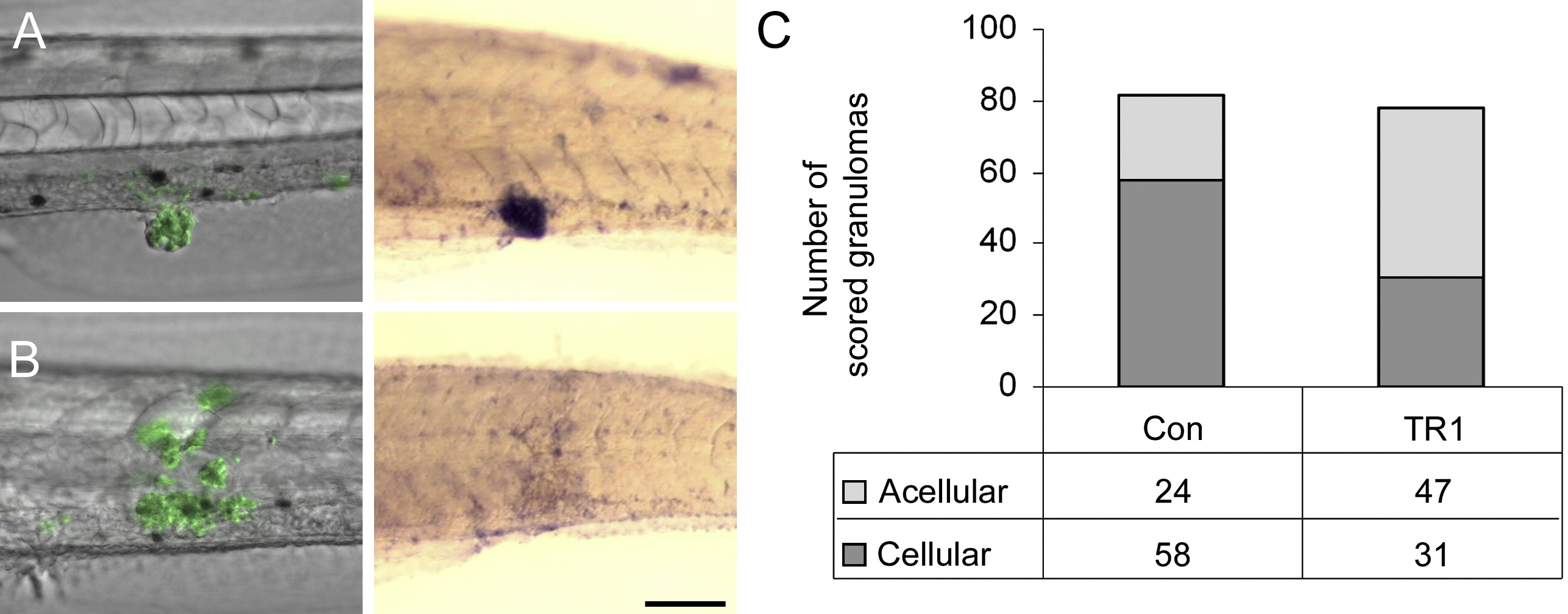
Fig. 4 TR1 Morphant Embryos Have Significantly More Acellular Granulomas than Controls
(A and B) Differential interference contrast overlay with fluorescence (left) of infected embryos then labeled with in situ hybridization for the macrophage marker fms (right). Green indicates M. marinum in fluorescent overlays. Purple staining of hybridized embryos indicates expression of the macrophage marker fms. (A) shows an example of a cellular granuloma in a control embryo before and after in situ hybridization indicating bacteria within aggregated macrophages. (B) shows an acellular granuloma example in a TR1 morphant embryo indicating bacteria found primarily outside of macrophages.
(C) The number of granulomas scored as either cellular or acellular in TR1 morphant versus control embryos. TR1 morphant granulomas were twice as likely to contain one-third or fewer fms-positive cells than to WT granulomas (p < 0.0001) as analyzed by Fisher's exact test of a contingency table. Embryos were 7 dpf and infected with 267 ± 28 CFU.
Reprinted from Immunity, 29(2), Clay, H., Volkman, H.E., and Ramakrishnan, L., Tumor necrosis factor signaling mediates resistance to mycobacteria by inhibiting bacterial growth and macrophage death, 283-294, Copyright (2008) with permission from Elsevier. Full text @ Immunity