
Fig. S1
- ID
- ZDB-IMAGE-080829-32
- Genes
- Publication
- England et al., 2006 - A dynamic fate map of the forebrain shows how vertebrate eyes form and explains two causes of cyclopia
- All Figures
- Figures for England et al., 2006
|
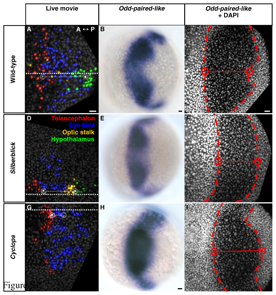
Fig. S1 Initiation of keel formation relative to eye-field location in wild-type, silberblick and cyclops embryos. (A-C) Wild type. (D-F) slb. (G-I) cyc morphant. (A-I) Dorsal views, anterior to left. (A,D,G) Forebrain regions, defined by cell tracking, at 80% epiboly (8.4 hpf). White cells and open circles - medial anterior eye (left) and initiation of keel formation (right). Adjoining solid line - intervening distance along the midline (square dotted line). Keel formation begins with hypothalamus in wild-type (A) and slb (D), and with anterior eye field in cyc morphant (G). For corresponding angular distances subtended at the embryonic centroid see Table S1. (B,E,H) Odd-paired-like (opl) gene expression, as determined using standard procedures, defines the eye field at 80% epiboly (8.4 hpf). Hypothalamus occupies the posterior-medial notch in wild type (B) and slb (E), consistent with the tracked data in A and D. However, eye tissue presides in the posterior-medial notch in cyc morphant (H). (C,F,I, Table S1) DAPI nuclear counter-staining, defining the medial boundaries (red cells and open circles) of the opl-expressing eye field (dashed line) at 80% epiboly (8.4 hpf), confirms this. hpf, hours post fertilisation. Scale bars: 25 μm.