
Fig. 5
- ID
- ZDB-IMAGE-080828-85
- Genes
- Antibodies
- Publication
- Kim et al., 2008 - Notch-regulated oligodendrocyte specification from radial glia in the spinal cord of zebrafish embryos
- All Figures
- Figures for Kim et al., 2008
|
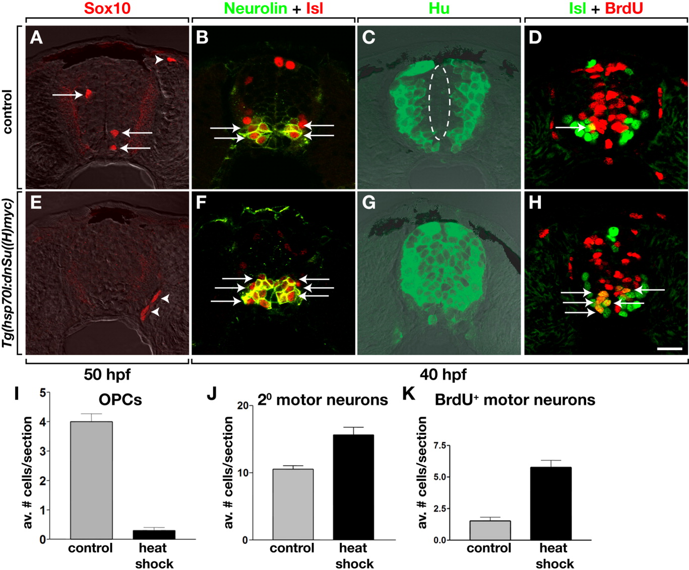
Fig. 5 Conditional inhibition of Notch signaling before OPC specification produces excess secondary motor neurons at the expense of OPCs. All images show transverse sections of spinal cord, dorsal to top. A-D: Control embryos. E-H: Tg(hsp70l:dnSu(H)myc) embryos heat-shocked at 30 hours postfertilization (hpf). Inhibition of Notch signaling before OPC specification blocks formation of Sox10+ OPCs (A,E,I) and produces excess secondary motor neurons (B,F,J). Arrows mark OPCs and secondary motor neurons and the small, dashed circle marks the area around the central canal, which is not occupied by motor neurons, in control embryos. The medial portion of the spinal cord, which does not have Hu+ neurons in control embryos (dashed circle, C) is filled with neurons in Notch-inhibited embryos (G). Pulse labeling with bromodeoxyuridine (BrdU, red) reveals that more dividing spinal cord cells adopt Isl+ motor neuron fates in Notch-inhibited embryos relative to controls (D,H,K). The quantitative data were obtained from sections of six control and six heat-shocked embryos for each experiment. Error bars represent SEM. Scale bar = 20 μM.