
|
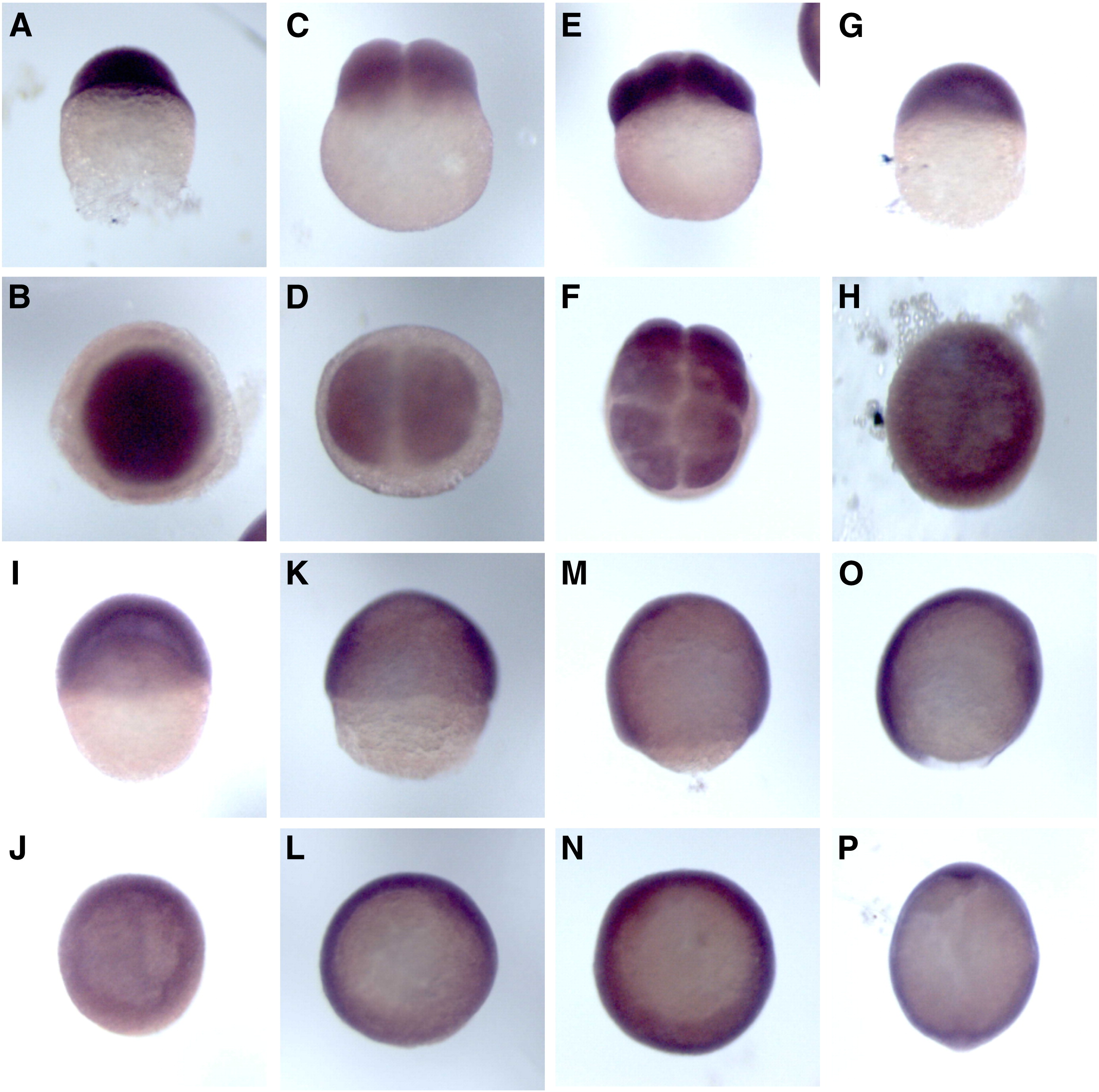
Fig. 3 Whole mount in situ hybridization of zLPTS at early stages of zebrafish embryos. Embryos were showed at different developmental stages such as 1-cell (0 hpf) (A and B), 2-cell (0.75 hpf) (C and D), 8-cell (1.25 hpf) (E and F), 30% epiboly (4.3 hpf) (G and H), ring stage (5.25 hpf) (I and J), 70% epiboly (7 hpf) (K and L), 90% epiboly (8.5 hpf) (M and N), 5 somites (12 hpf) (O and P). Hybridization was performed with a digoxigenin-labeled zLPTS antisense probe, and the purple reaction products were visualized by bright-field microscopy. Each pair is viewed from the side (A, C, E, G, I, K, M, O), top (B, D, F, H, J), or bottom (L, M and P).
Reprinted from Gene, 420(1), Sun, C., Wu, Z., Jia, F., Wang, Y., Li, T., and Zhao, M., Identification of zebrafish LPTS: A gene with similarities to human LPTS/PinX1 that inhibits telomerase activity, 90-98, Copyright (2008) with permission from Elsevier. Full text @ Gene