
Fig. 1
- ID
- ZDB-IMAGE-080828-6
- Genes
- Publication
- Dee et al., 2008 - Sox3 regulates both neural fate and differentiation in the zebrafish ectoderm
- All Figures
- Figures for Dee et al., 2008
|
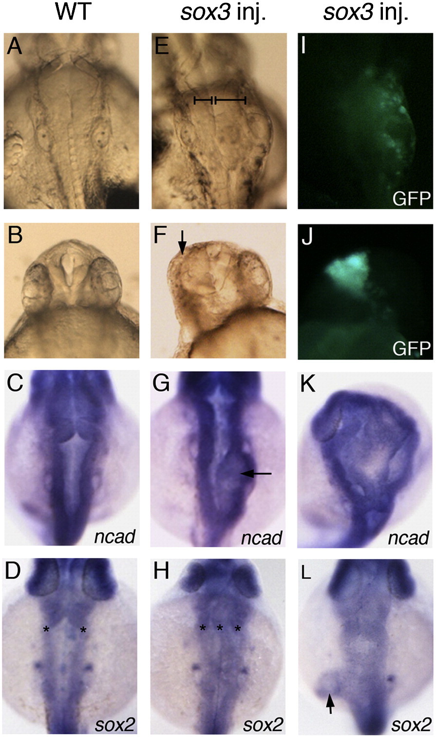
Fig. 1 Sox3 over-expression causes expansion of neural tissue. Wild-type control embryos (A–D) and embryos injected with sox3 mRNA at 16–32 cell stage (E–L) are viewed dorsally aged 24 hpf. (E, I) Injection of sox3 mRNA causes development of extra neural tissue visible in the live embryo (bars indicate unaffected and expanded sides of the CNS, E), and GFP fluorescence indicates this occurs in the region expressing exogenous sox3 (I). (F, J) Live embryo showing over-expression of sox3 causing loss of eye (arrow, F) when expressed in the rostral region of the CNS seen by GFP fluorescence (J). (C, D, G, H, K, L) In situ hybridisation for expression of neural markers ncad or sox2 in wild-type embryos (C, D) and embryos injected with sox3 mRNA (G, H, K, L) shows the development of ectopic tissue either within the brain (arrow, G; asterisks indicate multiple neural epithelia in panel H, 16/68 embryos), expansion of the brain (K, 2/21) or expansion of a local region of the trunk (arrow, L, 4/47).
Reprinted from Developmental Biology, 320(1), Dee, C.T., Hirst, C.S., Shih, Y.H., Tripathi, V.B., Patient, R.K., and Scotting, P.J., Sox3 regulates both neural fate and differentiation in the zebrafish ectoderm, 289-301, Copyright (2008) with permission from Elsevier. Full text @ Dev. Biol.