Image
Figure Caption
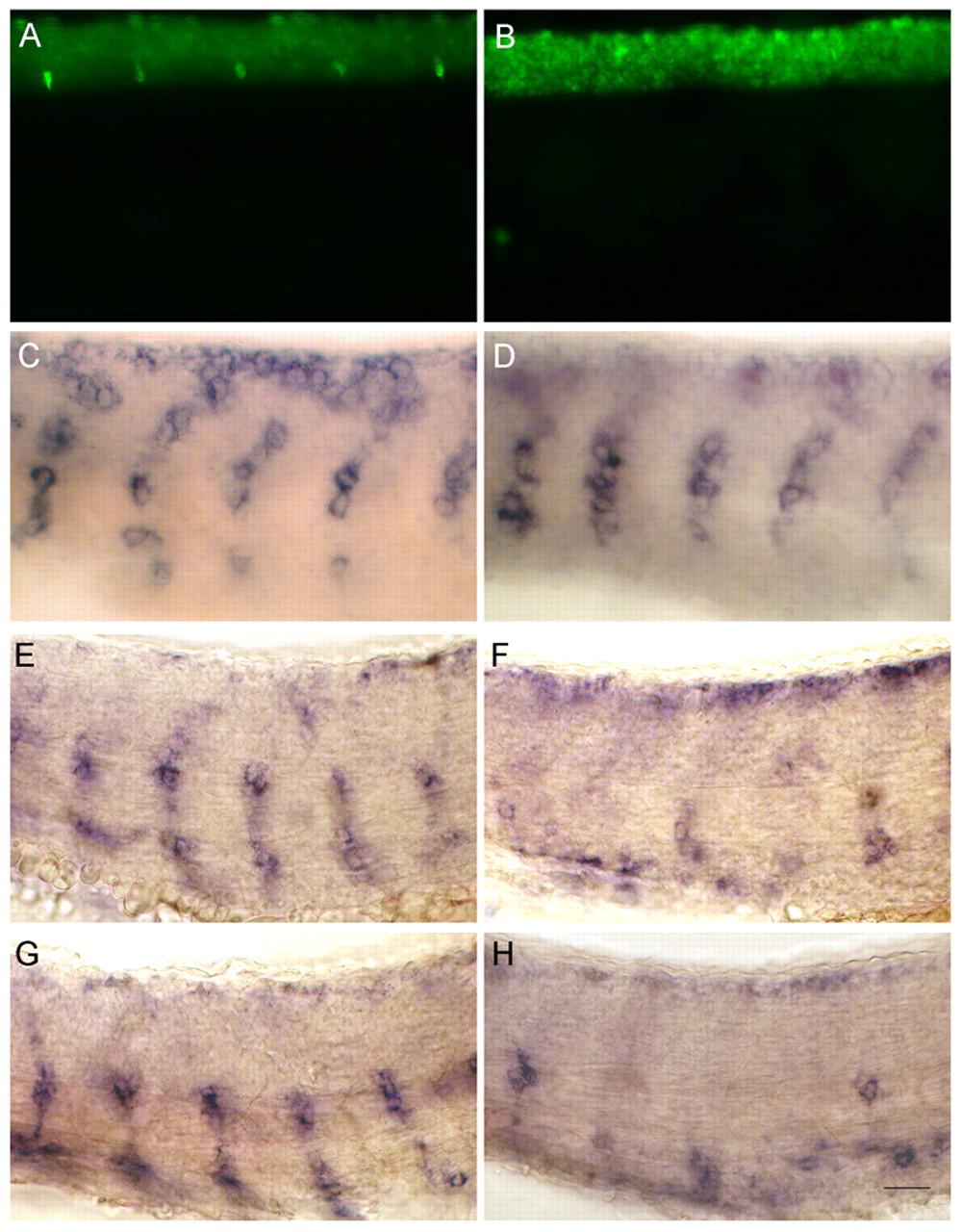
Fig. 8 nrg1 and nrg2a act redundantly during DRG neuron formation. (A,B) Elav1 antibody labeling at 4 dpf. Co-injection of nrg1 and nrg2a MOs resulted in absence of DRG neurons (B), whereas DRG neurons are normal in controls (A). (C-H) Whole-mount wild-type (C,E,G) and nrg1 plus nrg2a MO-injected embryos (D,F,H) labeled with crestin riboprobe at 24 hpf (C,D), 27 hpf (E,F) and 30 hpf (G,H). Like erbb3b and erbb2 mutants, double MO-injected embryos have normal early NC migration (C,D) but, at later stages, NC migration is disrupted (E-H). Scale bar: 20 μm.
Figure Data
Acknowledgments
This image is the copyrighted work of the attributed author or publisher, and
ZFIN has permission only to display this image to its users.
Additional permissions should be obtained from the applicable author or publisher of the image.
Full text @ Development

