Image


                        Figure Caption


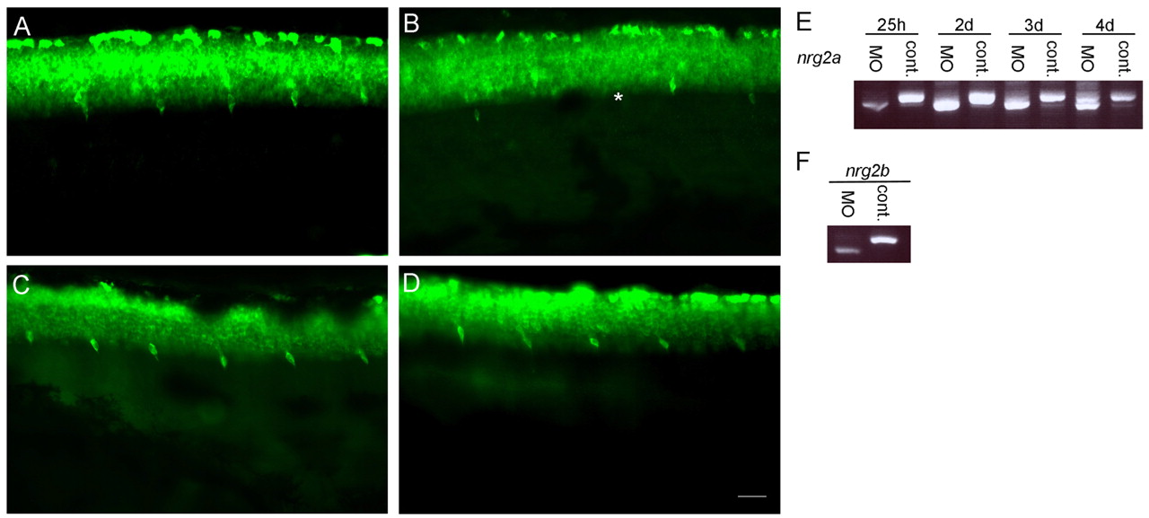
Fig. 7 nrg2a alone is unnecessary for DRG neuron formation. DRG neurons labeled with Elavl antibody at 4 dpf. DRG neurons in controls (A,C). DRG neurons are missing from some segments of nrg2a MO-injected embryos (B) but appear normal in nrg2b MO-injected embryos (D). Asterisks indicate absent DRG neurons in B. (E) RT-PCR showing that nrg2a MOs cause incorrect mRNA splicing through 3 dpf, but by 4 dpf, both incorrectly-spliced and correctly-spliced transcripts are present. (F) RT-PCR showing that both the nrg2a and nrg2b MOs cause incorrect splicing at 25 hpf. Scale bar: 20 μm.
                
                    
                        Figure Data


                        Acknowledgments


This image is the copyrighted work of the attributed author or publisher, and
ZFIN has permission only to display this image to its users.
Additional permissions should be obtained from the applicable author or publisher of the image.
      Full text @ Development


        