
|
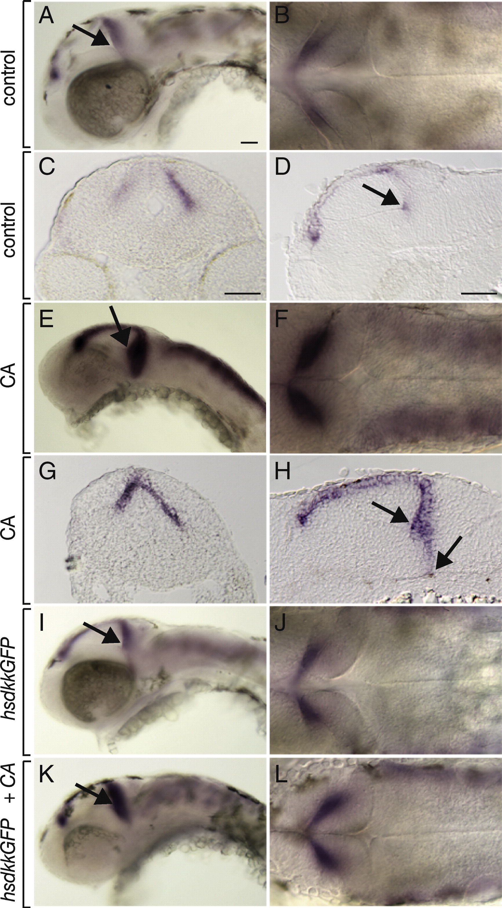
Fig. 7 Inhibition of Hh signaling causes expansion of the wnt1 expression domain. All embryos shown are 48 hpf. Black arrows in whole mount views depict the morphological line of the midbrain adjoining the midbrain–hindbrain boundary. Lateral whole mount (A), dorsal whole mount (B), transverse section (C) and sagittal section (D) showing wnt1 expression in control embryos. The black arrows mark the base of the 3rd ventricle, with which the ventral extent of wnt1 expression coincides in the midbrain–hindbrain region. (E–H) Similar views of wnt1 expression in embryos treated with CA beginning at 6 hpf. wnt1 expression in the midbrain–hindbrain region is expanded both ventrally and posteriorly. (I, J) Expression of wnt1 in embryos that overexpressed Dkk1 at 30 hpf was similar to controls. (K, L) wnt1 expression was expanded in embryos treated with CA at 6 hpf and heat shocked to induce Dkk1 expression at 30 hpf. Scale bars represent 20 μm in all panels.
Reprinted from Developmental Biology, 318(1), McFarland, K.A., Topczewska, J.M., Weidinger, G., Dorsky, R.I., and Appel, B., Hh and Wnt signaling regulate formation of olig2(+) neurons in the zebrafish cerebellum, 162-171, Copyright (2008) with permission from Elsevier. Full text @ Dev. Biol.