
Fig. 4
- ID
- ZDB-IMAGE-080617-9
- Genes
- Antibodies
- Publication
- Anderson et al., 2008 - Loss of unc45a precipitates arteriovenous shunting in the aortic arches
- All Figures
- Figures for Anderson et al., 2008
|
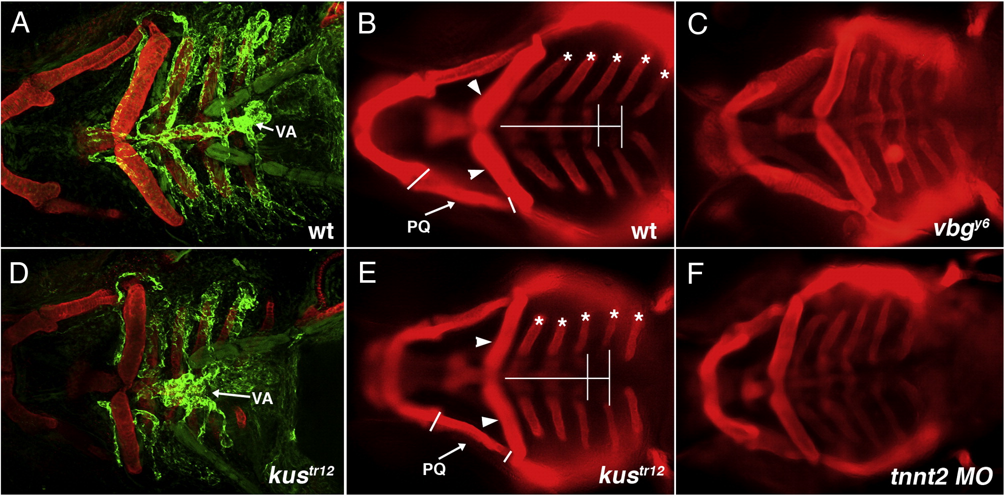
Fig. 4 Increased distance between left and right posterior ceratobranchial cartilages is a specific kustr12 phenotype. Aortic arches (bright green) develop in close association with pharyngeal cartilages (red) in WT (A) and kustr12 (D). Note the malformed ventral aorta (VA) in kustr12. (The darker green staining is background and represents pharyngeal muscle.) Defects in jaw and pharyngeal cartilages in kustr12 include shortening of the palatoquadrates (PQ); compression of the basibranchial cartilages (horizontal white line); improper angling of the ceratobranchial (asterisks) and ceratohyal (arrowheads) cartilages; and decreased lateral distance (vertical white lines) between left and right ceratobranchial cartilages in pharyngeal arches 5 and 6 [compare B (WT) to E (kustr12)]. Like kustr12, vbgy6 mutants (C) and tnnt2 morphants (F) develop edema and exhibit all described cartilage defects except increased lateral distance between posterior ceratobranchials. Thus, only the latter defect is likely a direct effect of the kustr12 mutation. A, D: 2D confocal projections of TG(flk1:GFPla116; gata1:dsRed) embryos stained with anti-collagen II (red) and anti-GFP (green). B, C, E, F: Macro images of embryos stained with anti-collagen II. All embryos are 4 dpf; ventral views, anterior to the left.
Reprinted from Developmental Biology, 318(2), Anderson, M.J., Pham, V.N., Vogel, A.M., Weinstein, B.M., and Roman, B.L., Loss of unc45a precipitates arteriovenous shunting in the aortic arches, 258-267, Copyright (2008) with permission from Elsevier. Full text @ Dev. Biol.