
IMAGE
Fig. 2
- ID
- ZDB-IMAGE-080613-22
- Genes
- Publication
- Toyama et al., 2008 - Brd4 associates with mitotic chromosomes throughout early zebrafish embryogenesis
- All Figures
- Figures for Toyama et al., 2008
Image
|
Figure Caption
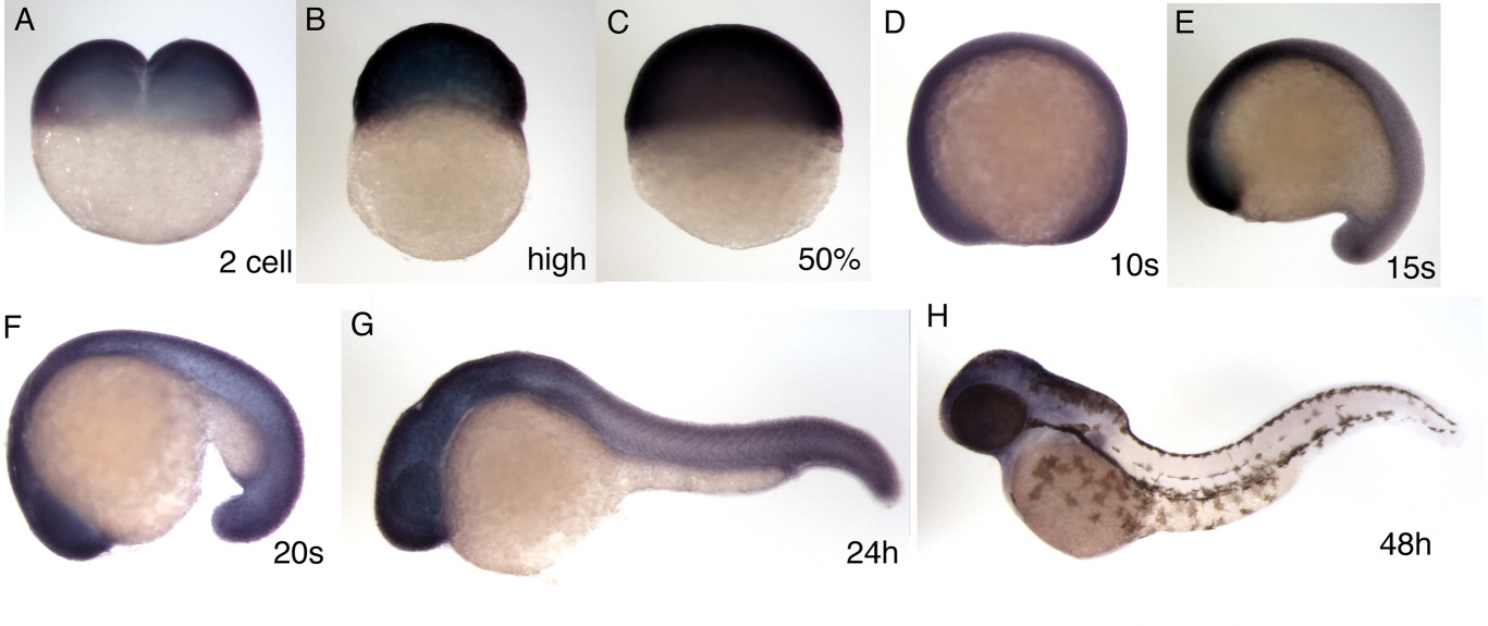
Fig. 2 Expression pattern of the zebrafish brd4 gene. A-H: The stage examined by in situ hybridization, carried out as described by Toyama and Dawid ([1997]), is shown in each panel: two-cell stage (A), high stage (B), 50% epiboly (C), 10 somite (D), 15 somite (E), 20 somite (F), 24 hours postfertilization (hpf; G) and 48 hpf (H). D-H: All lateral view, anterior is left.
Figure Data
Acknowledgments
This image is the copyrighted work of the attributed author or publisher, and
ZFIN has permission only to display this image to its users.
Additional permissions should be obtained from the applicable author or publisher of the image.
Full text @ Dev. Dyn.