
Fig. 1
- ID
- ZDB-IMAGE-080529-58
- Genes
- Publication
- Gregg et al., 2003 - Positional cloning of the young mutation identifies an essential role for the Brahma chromatin remodeling complex in mediating retinal cell differentiation
- All Figures
- Figures for Gregg et al., 2003
|
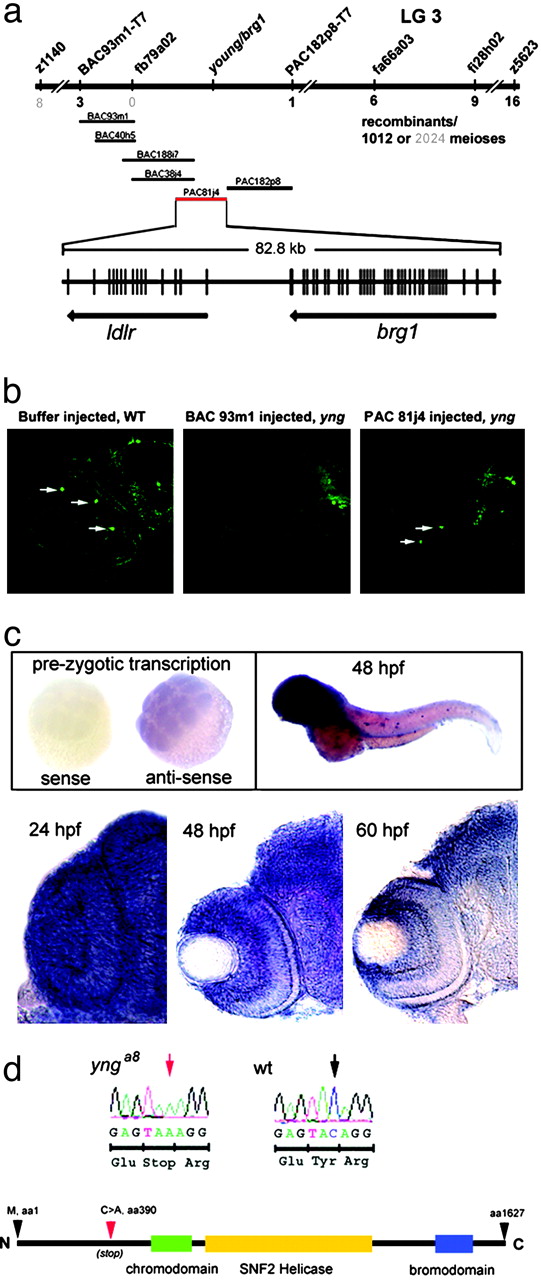
Fig. 1 yng is caused by a mutation in brg1. (a) Genetic and physical map. Linkage was initially discovered between simple sequence length polymorphism markers z1140 and z5623. Polymorphisms within ESTs mapped to this region on the T51 radiation hybrid panel and were then used to refine the interval. EST fb79a02 was found to contain no recombinants of 2,024 meioses, and BAC and PAC clones were used to build a contig around this locus. PACs 182p8 and 81j4 and BAC 38j4 were sequenced to reveal a high level of synteny with human chromosome 19 based on BLAST results. Positive rescue experiments using PAC 81j4 limited the search to either zebrafish ldlr or brg1. Vertical lines are exons determined by PCR of embryonic cDNA and based on GENSCAN predictions. (b) BAC/PAC rescue analysis. Transverse retinal sections showing the presence of TH (a late interplexiform cell marker, arrows) in a WT embryo injected with control buffer, a yng mutant injected with BAC 93m1 (which does not contain brg1 and is negative for TH expression), and a yng mutant injected with PAC 81j4 (which does contain brg1 and rescued TH expression). Arrows indicate retinal TH-positive neurons. TH is also normally expressed in the ventral forebrain of both yng and WT embryos (Right). (c) In situ mRNA analysis of brg1. Sixteen-cell stage embryos, a time before zygotic transcription begins, shows that brg1 transcript is provided maternally. Results with sense control and antisense riboprobes are shown. Whole-mount analysis at 48 hpf reveals strong anterior brg1 expression. Transverse retinal sections at 24, 48, and 60 hpf show robust brg1 expression during differentiation. (d) Sequence analysis and predicted protein structure. A C→ A transversion was identified in cDNA from yng homozygous mutants and later confirmed through genomic sequence analysis (red arrow). This base substitution changes a tyrosine codon to a stop codon very early with in the coding sequence of brg1 (red arrowhead). This truncation deletes all of the identified functional domains.