
Fig. 5
- ID
- ZDB-IMAGE-080505-5
- Publication
- Snelson et al., 2008 - Tbx2b is required for the development of the parapineal organ
- All Figures
- Figures for Snelson et al., 2008
|
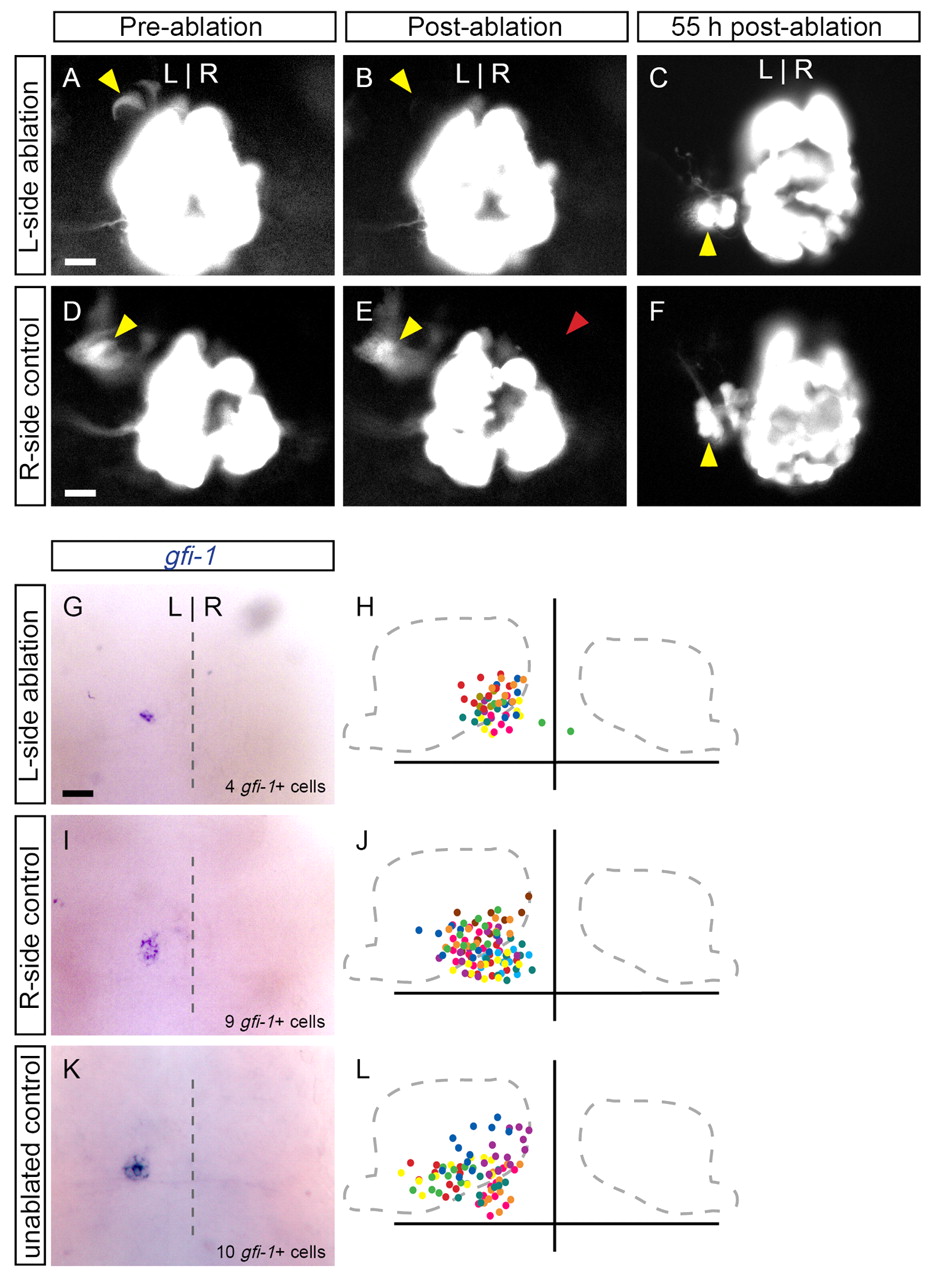
Fig. 5 Reduced numbers of parapineal cells are able to migrate to the left side of the brain. Dorsal views of the epithalamus in (A,B,D,E) 31-hpf zebrafish embryos or (C,F,G-L) 55-hpf larvae. Expression of foxd3:gfp labels parapineal precursor cells (A, yellow arrowhead), which are subsequently ablated with laser pulses (B). The remaining parapineal cells (average of 5±2 cells, n=18) migrate to the left side of the brain, as revealed by foxd3:gfp (C) and gfi1 (G) expression. In H, the position of gfi1-expressing cells in ten larvae are overlaid, with different colors representing individual samples. (D-F) As a control, cells contralateral to the parapineal precurors were ablated (red arrowhead). The number of parapineal cells (10±1, n=8) and their migration in these controls (I,J) were identical to those in unablated controls (K,L; 10±2 cells, n=10). Scale bars: 25 μm.