
|
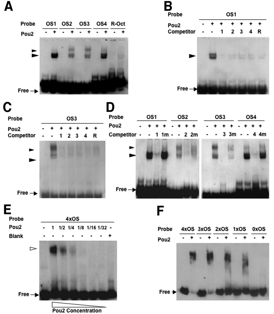
Fig. 8 Binding activities of the octamer sequences in the pou2 regulatory region with the Pou2 protein. A: Digoxigenin (DIG) -labeled probes for the four octamer sequences (OS1-4) and R-Oct sequence generated shifted bands in the presence of Pou2. The fast-migrating doublet bands marked with large arrowheads were generated by all the probes, whereas the slower-migrating bands, shown with small arrowheads, were seen only when the pou2 octamer sequences were used. B,C: Binding of Pou2 with DIG-labeled OS1 (B) and OS3 (C) was competed out with 100-fold molar excess of the unlabeled oligos (OS1-4 and R-Oct). D: Binding of Pou2 with the four octamer probes was competed with 100-fold excess of cognate oligos (OS1-4), but not at all or only partially by mutated oligos (OS1m-4m). E: Binding of Pou2 with 4×OS DNA containing the four octamer sequences (OS1-4) generated a single shifted band with no intermediate bands. F: Binding of Pou2 with 4xOS, 3xOS, 2xOS, 1xOS, and 0xOS, which included decreasing numbers of the OS sequences, was examined, showing a gradual decrease in size of the complex and an increase in the amount of free probes.