
IMAGE
Fig. 3
- ID
- ZDB-IMAGE-080411-64
- Antibodies
- Publication
- Wang et al., 2008 - Inactivation of zebrafish mrf4 leads to myofibril misalignment and motor axon growth disorganization
- All Figures
- Figures for Wang et al., 2008
Image
|
Figure Caption
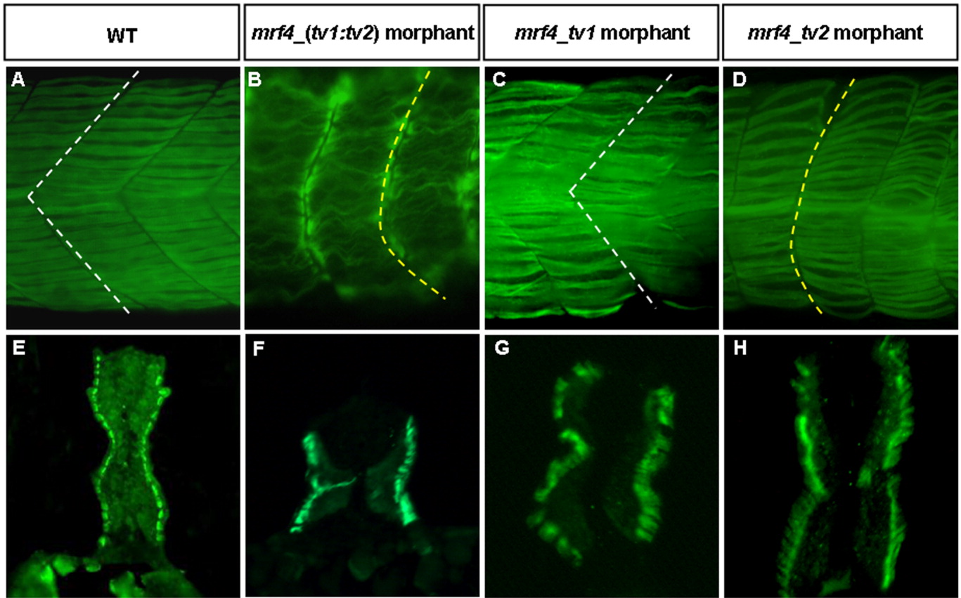
Fig. 3 F59 monoclonal antibody-staining of the 36-hpf of WT zebrafish embryos (A; cross section at E), mrf4_(tv1:tv2)-morphants (B,F), mrf4_tv1-morphants (C,G), and mrf4_tv2-morphants (D,H). White dashed lines in A and C indicate the boundaries of chevron-shaped somites. Yellow dashed lines in B and D indicate the boundaries of U-shaped somites.
Figure Data
Acknowledgments
This image is the copyrighted work of the attributed author or publisher, and
ZFIN has permission only to display this image to its users.
Additional permissions should be obtained from the applicable author or publisher of the image.
Full text @ Dev. Dyn.