
Fig. 6
- ID
- ZDB-IMAGE-080411-6
- Genes
- Publication
- Wolman et al., 2008 - Transient axonal glycoprotein-1 (TAG-1) and laminin-alpha1 regulate dynamic growth cone behaviors and initial axon direction in vivo
- All Figures
- Figures for Wolman et al., 2008
|
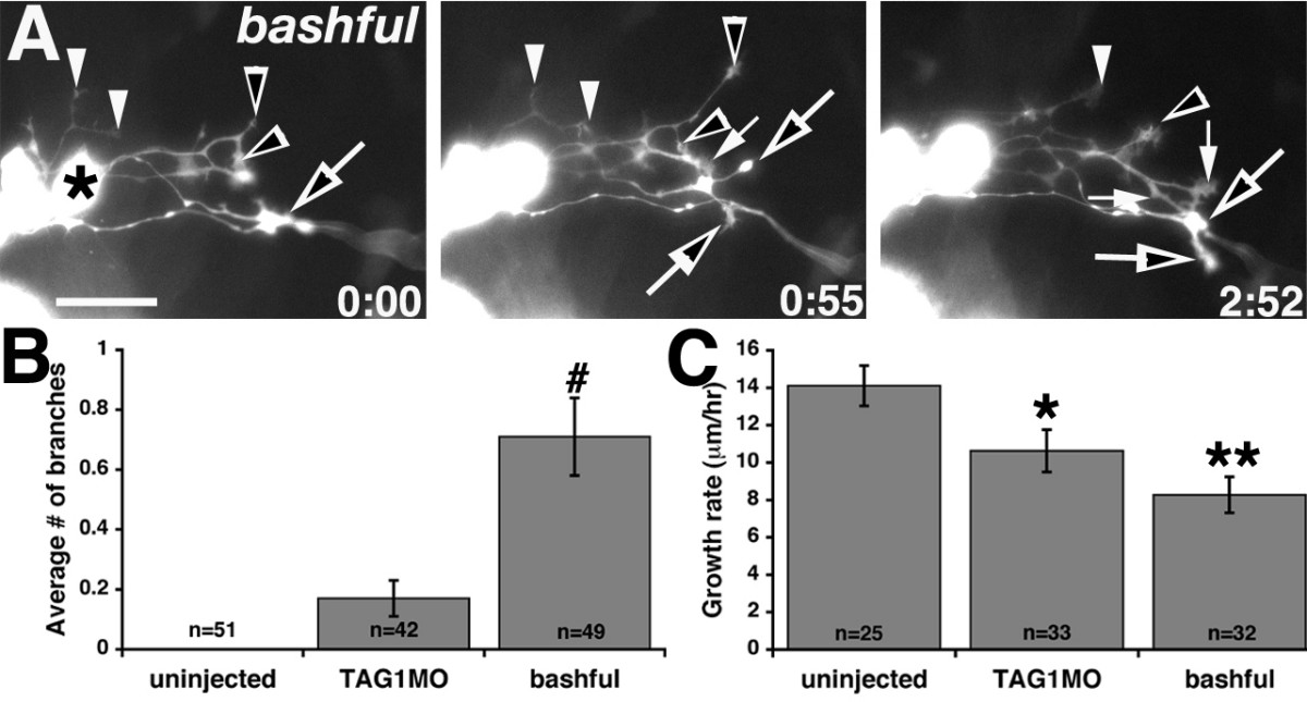
Fig. 6 bal embryos have excessively branched axons and MLF growth rates are reduced by TAG-1 or laminin-α1 loss of function.(a) Images from timelapse sequence of MLF axon outgrowth in a bal;Tg(pitx2c:gfp) embryo. Ventral views, anterior to the left, midline is up. The asterisk denotes the caudal-most nucMLF cell. Open and filled arrows and arrowheads label branches from individual axons. The time stamp shows hours: minutes. (b, c) Quantification of the average number of branches per axon (b) and the average growth rate of MLF axons (c). #P < 0.001, Kruskal-Wallis ANOVA versus uninjected. *P < 0.05, **P < 0.001, two-tailed t-test versus uninjected. Error bars represent the standard error of the mean. N equals the number of axons. Scale bar = 25 μm.