
Fig. 2
- ID
- ZDB-IMAGE-080411-16
- Genes
- Publication
- Insinna et al., 2008 - The homodimeric kinesin, Kif17, is essential for vertebrate photoreceptor sensory outer segment development
- All Figures
- Figures for Insinna et al., 2008
|
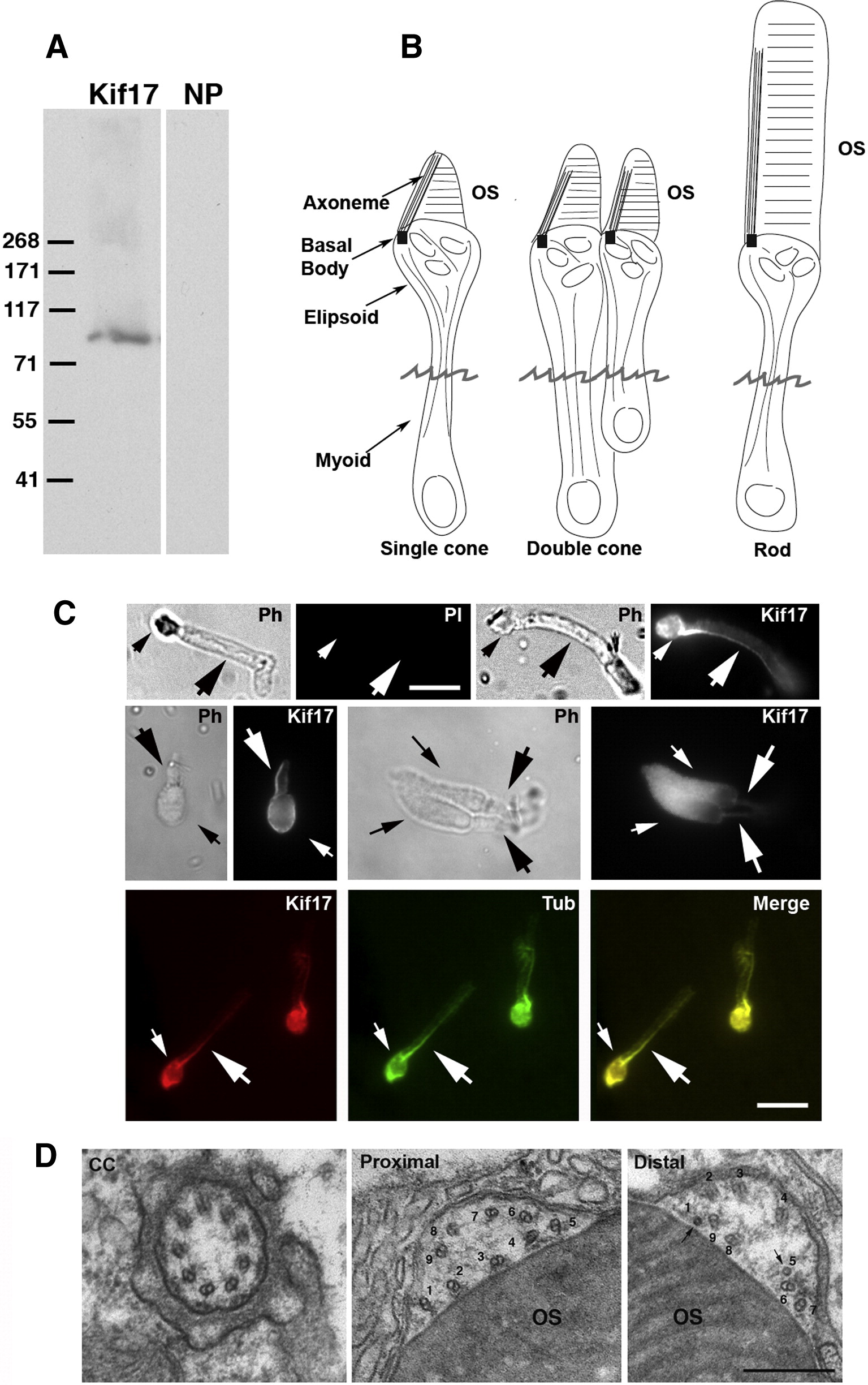
Fig. 2 Immunocytochemical (IC) localization of Kif17 in photoreceptors. (A) Immunoblot of 3-day zebrafish embryo lysate. The Kif17 band corresponds to the predicted size based on the zebrafish cDNA sequence; NP, no primary antibody. (B) Three morphological types of zebrafish photoreceptors; isolated photoreceptors break off at the position of the jagged line. (C) IC of Kif17 in isolated adult zebrafish photoreceptors. Ph, Upper left is a phase (PH), and no primary antibody control image (PI) of a rod. Upper right is a phase and a Kif17 stained rod. Middle panel is a phase and Kif17 stained single cone (left), and double cone (right). Lower panel shows co-localization of Kif17 (left) with acetylated α-tubulin (middle) along the axoneme of a rod. Small arrows point to the ellipsoid region and the large arrows point to axoneme in the OS. Bar = 10 μm. (D) EM images at the level of the connecting cilium (CC), the proximal OS, and more distal OS of a developing cone from a 3-day zebrafish larva. Note that both the proximal and distal OS images are within the most proximal one-third of the OS. Doublets adjacent to the discs are numbered 1–9; the more distal image shows two singlets (1 and 5) and 7 doublets adjacent to the OS discs. Bar = 0.33 μm.
Reprinted from Developmental Biology, 316(1), Insinna, C., Pathak, N., Perkins, B., Drummond, I., and Besharse, J.C., The homodimeric kinesin, Kif17, is essential for vertebrate photoreceptor sensory outer segment development, 160-170, Copyright (2008) with permission from Elsevier. Full text @ Dev. Biol.