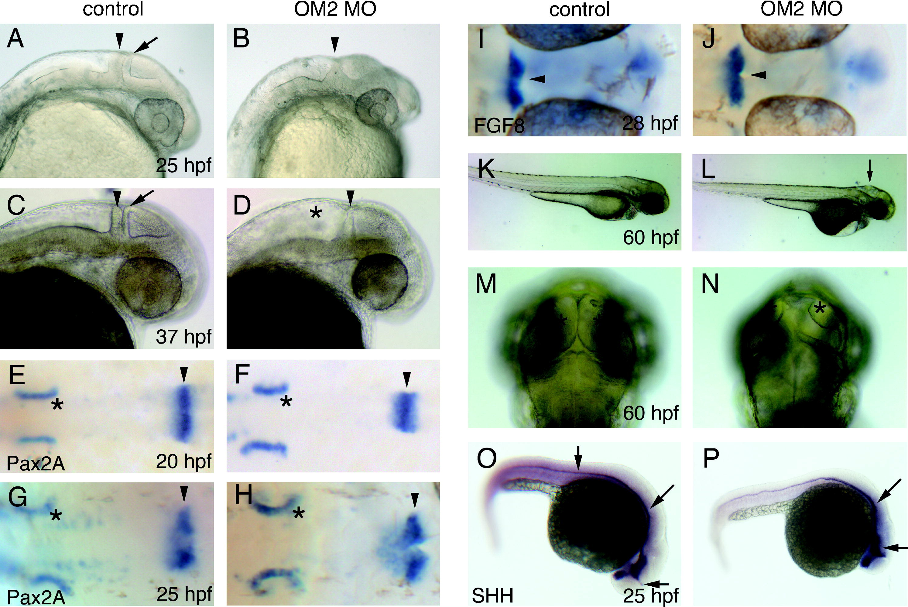
Fig. 3 OM2 morphants show specific defects in anterior CNS development (n = 78). (A and B) Embryos (25 hpf) from the lateral view show specific defects of OM2 morphants in the midbrain–hindbrain boundary (MHB), which appears to have been formed by the fusion of tectal (arrow) and cerebellar walls (arrowheads). Slightly smaller size of the morphant eyes is also shown. (C and D) At 37 hpf, the MHB defect is still present. (E–H) Pax2a in situ hybridization patterns are not affected by OM2 knockdown. Asterisks indicate otic vesicles. Pax2a mRNA expression at the MHB is intact in OM2 morphants (arrowheads), despite the morphological defects (B and D). (I and J) MHB-specific Fgf8 mRNA expression pattern is not altered by OM2 knockdown. Arrowheads indicate Fgf8 mRNA at the MHB. (K and L) At 60 hpf, gross morphology of OM2 morphants is similar to that of control MO injected fish, except for mild edema in the 4th ventricle (arrow in L). (M and N) Dorsal side of the head shows markedly decreased size of the optic tectum (asterisks). Notice also that the MHB is significantly thinner and less developed than its control counterpart. (O and P) In situ hybridization with SHH probes shows no alteration in SHH mRNA expression patterns in OM2 morphants (arrows; n = 24).
Reprinted from Mechanisms of Development, 125(1-2), Lee, J.A., Anholt, R.R., and Cole, G.J., Olfactomedin-2 mediates development of the anterior central nervous system and head structures in zebrafish, 167-181, Copyright (2008) with permission from Elsevier. Full text @ Mech. Dev.

