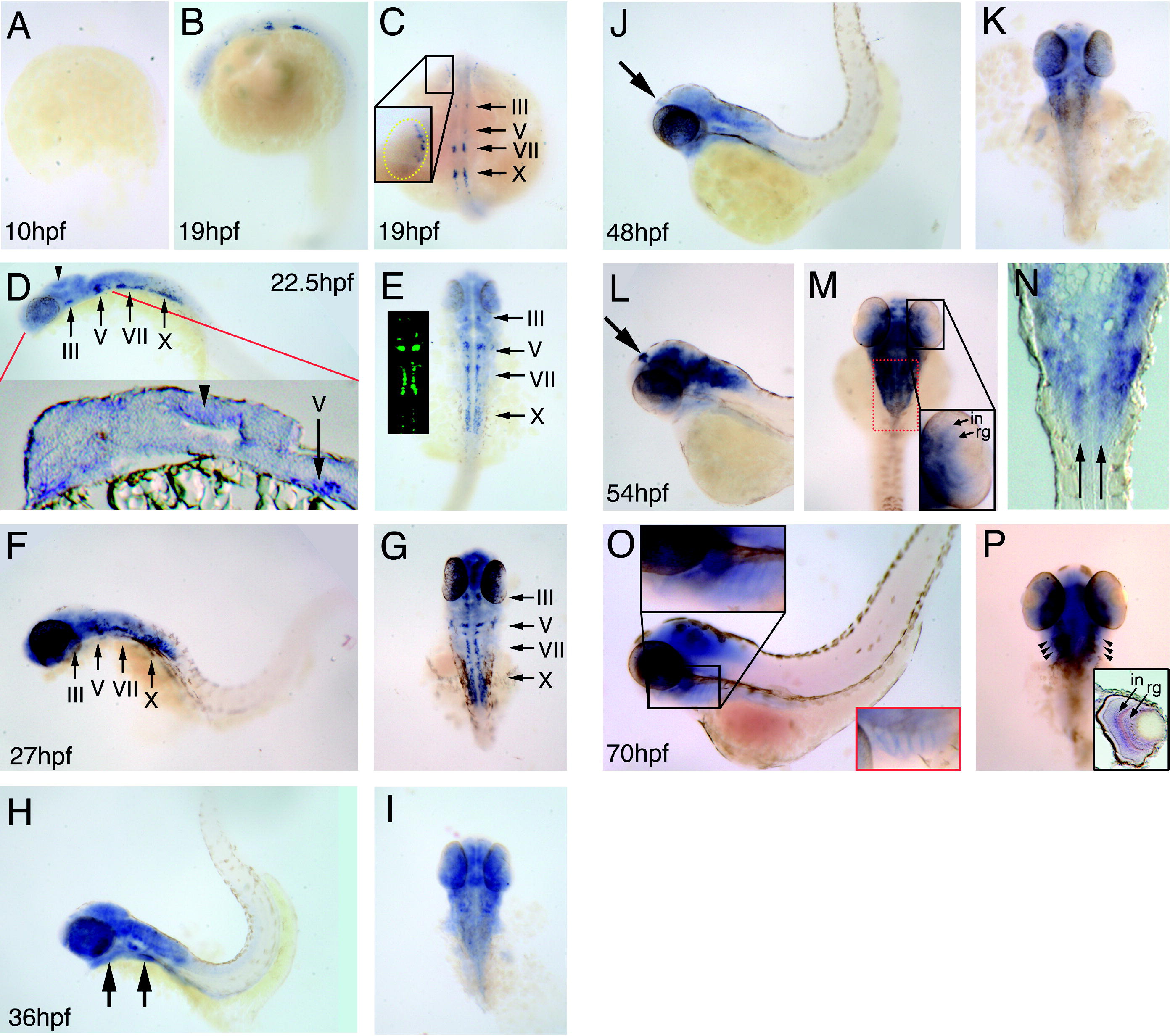
Fig. 2 Localization of OM2 mRNA in developing zebrafish using in situ hybridization. (A) OM2 mRNA is not detected in 10 hpf embryos. (B and C) Embryo (19 hpf) shows OM2 mRNA in the developing eye and branchiomotor nuclei (III, V, VII and X). Expression in the proximal region of the optic primordium is shown in the inset (yellow dotted line shows the boundary of optic primordium). (D and E) In 22.5 hpf embryos broader domains of OM2 mRNA expression were detected as well as the sharply defined branchiomotor nuclei-specific expression. GFP expression in branchiomotor nuclei of an islet-GFP embryo is also shown in (E) demonstrating the branchiomotor identity of these nuclei. Arrowhead in (D) indicates a strong expression of OM2 in the tectum, which is also observed in a cryostat section (arrowhead in the inset). (F and G) OM2 expression pattern in a 27 hpf embryo. Strong expression is detected in the forebrain and the tectum as shown in G. (H and I) Embryo (36 hpf) shows strong expression of OM2 mRNA in the anterior CNS, including ventral regions that will develop into the lower jaw and pharyngeal arches (arrows in H). In addition, OM2 is clearly detected in the eyes. (J and K) Embryos (48 hpf), showing OM2 expression in eye and brain, the epiphysis (arrow in J), with branchiomotor nuclei expression still being observed (J). (L and M) In 54 hpf embryos OM2 expression in forebrain, tectum, epiphysis and eyes is detected. More stratified patterns of OM2 mRNA are observed in the inner nuclear cell layer (in) and retinal ganglion cell layer (rg) in the eye (M). Close observation with cryosections shows that OM2 mRNA is still expressed in vagal (CN X) branchiomotor nuclei (arrows in N: red dotted box in M indicates the region shown in cryosection N). OM2 in developing pharyngeal arches is more restricted and this pattern is continued to 70 hpf embryos. (O and P) Embryo (70 hpf) shows a similar pattern to 54 hpf embryos. Notice a strong, yet highly restricted pattern of OM2 mRNA expression is found along the pharyngeal arches (red inset in O was obtained from an oblique angle). Also in cryosections of 70 hpf embryonic eye OM2 mRNA is expressed in defined, laminated layers of the INL and RGC, as in 54 hpf embryos (inset in P). Small arrowheads in P indicates OM2 mRNA expression in pharyngeal arches.
Reprinted from Mechanisms of Development, 125(1-2), Lee, J.A., Anholt, R.R., and Cole, G.J., Olfactomedin-2 mediates development of the anterior central nervous system and head structures in zebrafish, 167-181, Copyright (2008) with permission from Elsevier. Full text @ Mech. Dev.

