
Image
|
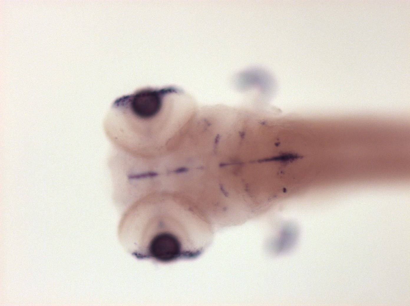
Figure Caption
Fig. 7
Expressed in brain ventricular zone, in optic nerve, in pectoral fin epidermis and in cornea
Please note that in 5 day old embryos some structures are not accessible to the probe (such as notochord, most of the trunk and tail). Therefore the description of the expression pattern is only partial.
Orientation
| Preparation | Image Form | View | Direction |
| whole-mount | still | dorsal | anterior to left |
Figure Data