
Image
|
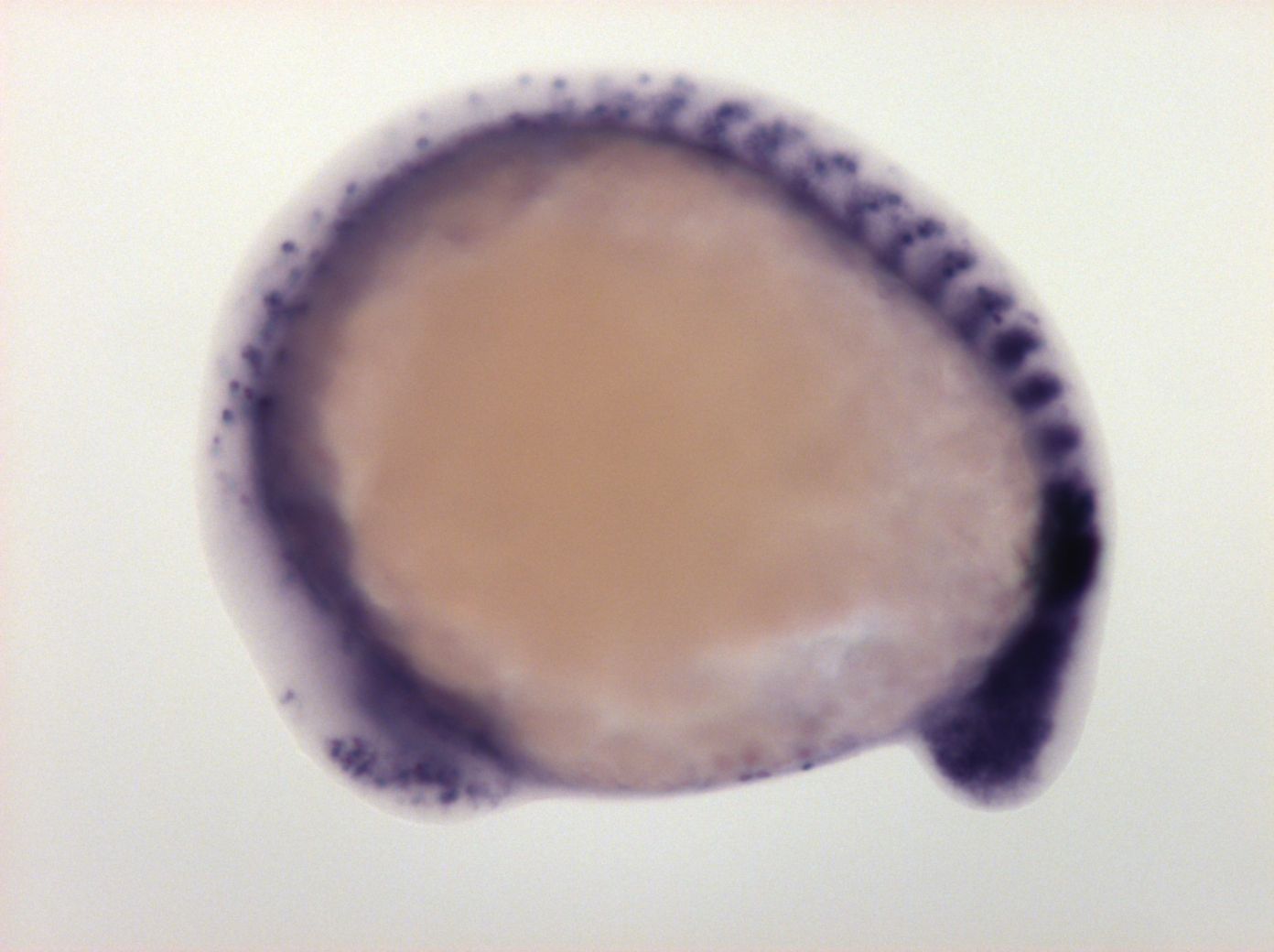
Figure Caption
Fig. 3 posterior part of somites, last formed somites, weak in unsegmented paraxial mesoderm, hypochord (or endoderm), ventral and dorsal neurons, mucus secreting cell, posterior telencephalon, anterior diencephalon, retina
Orientation
| Preparation | Image Form | View | Direction |
| whole-mount | still | side view | anterior to left |
Figure Data