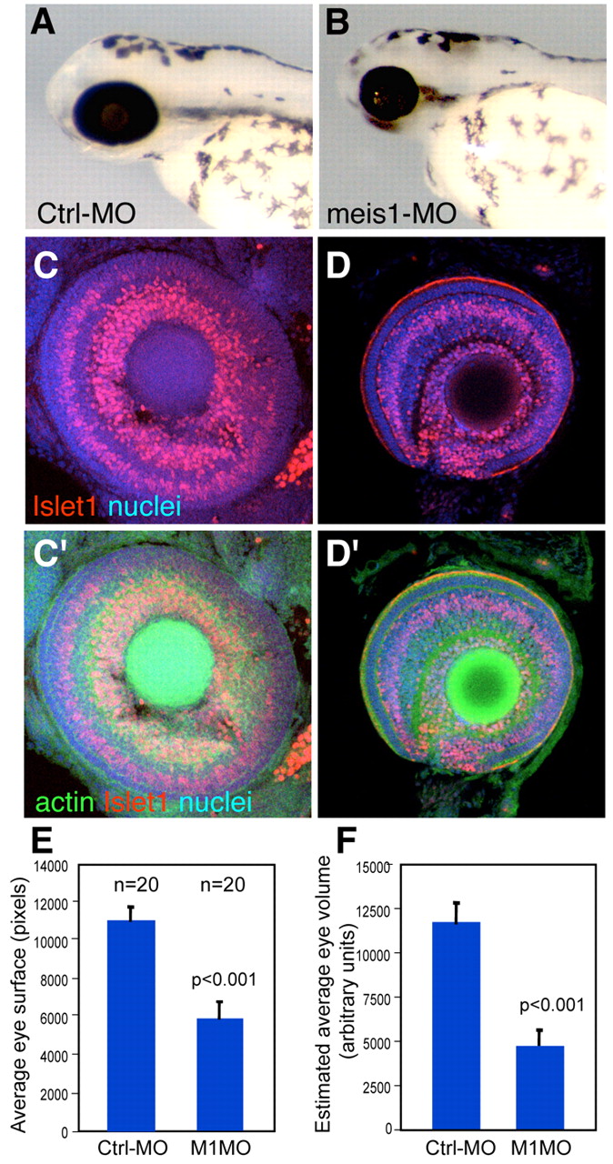
Fig. 2 meis1 is required for the growth of the eye primordium. (A,B) Lateral views of representative 72hpf control-MO (A) and meis1-MO (B) -injected fish. meis1 morphants are microphthalmic. (C,D) Confocal images of dissected eyes stained for propidium iodide (nuclei), rhodamine-phalloidin (filamentous actin) and Islet1, which labels GCL nuclei and some in the INL. The reduced eyes from meis1 morphants show apparently normal retina lamination (D,D′), but fewer cells than control eyes (C,C'). Area (E) and estimated volume (F) of control-MO and meis1-MO-injected embryos at 72hpf. meis1- morphant embryos show a significant (P<0.001) reduction in eye area and volume (45% and 60%, respectively). n=20 for each condition.
Image
Figure Caption
Figure Data
Acknowledgments
This image is the copyrighted work of the attributed author or publisher, and
ZFIN has permission only to display this image to its users.
Additional permissions should be obtained from the applicable author or publisher of the image.
Full text @ Development

