
IMAGE
Fig. 3
- ID
- ZDB-IMAGE-080325-16
- Publication
- Hendricks et al., 2008 - Disruption of Esrom and Ryk identifies the roof plate boundary as an intermediate target for commissure formation
- All Figures
- Figures for Hendricks et al., 2008
Image
|
Figure Caption
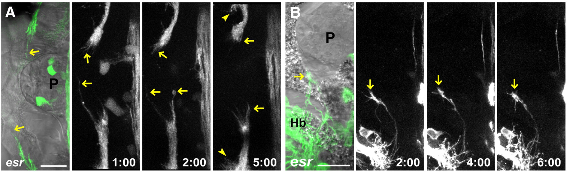
Fig. 3 Mutant axons stall at the roof plate boundary. (A) Time series in an esrom;Kaede embryo shows axons oriented toward the midline and extending and retracting (arrows, see Supplementary Movie 3). After several hours, innervation can be seen developing in the ipsilateral neuropil (arrowhead). (B) Time series of a single mutant growth cone (arrow), maintaining a complex morphology and dynamic behavior for several hours (see Supplementary Movie 4). P: pineal. Scale bar = 20 μm.
Acknowledgments
This image is the copyrighted work of the attributed author or publisher, and
ZFIN has permission only to display this image to its users.
Additional permissions should be obtained from the applicable author or publisher of the image.
Reprinted from Molecular and cellular neurosciences, 37(2), Hendricks, M., Mathuru, A.S., Wang, H., Silander, O., Kee, M.Z., and Jesuthasan, S., Disruption of Esrom and Ryk identifies the roof plate boundary as an intermediate target for commissure formation, 271-283, Copyright (2008) with permission from Elsevier. Full text @ Mol. Cell Neurosci.