
Fig. 5
- ID
- ZDB-IMAGE-080305-3
- Publication
- Sato et al., 2005 - Mutually exclusive glomerular innervation by two distinct types of olfactory sensory neurons revealed in transgenic zebrafish
- All Figures
- Figures for Sato et al., 2005
|
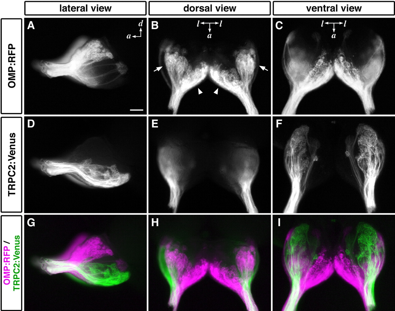
Fig. 5 Differential axonal projections from two types of OSNs. Whole-mount OB from OMP:RFP;TRPC2:Venus adult zebrafish were observed under a fluorescence stereomicroscope: A, D, G, lateral views; B, E, H, dorsal views; C, F, I, ventral views. Ciliated OSNs labeled with RFP projected their axons to almost all over the dorsal region and the ventromedial portion of the OB (A-C; magenta in G-I). In contrast, microvillous OSNs labeled with Venus projected their axons exclusively to the ventrolateral region (D-F; green in G-I). The arrows and arrowheads indicate dorsal cluster and the anterior plexus, respectively. a, Anterior; d, dorsal; l, lateral. Scale bar, 100 μm.