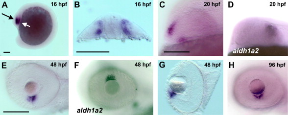
Fig. 2 Expression of aldh1a3 in the developing eye. Expression of aldh1a3 (A–C, E, G, H) and aldh1a2 (D and F) was detected by whole mount in situ hybridisation in the zebrafish eye. aldh1a3 was first detected in the eye primordium at 12–16 hpf (not shown; A, black arrow; B, transverse section through the plane indicated by the white arrow in A). While aldh1a3 is expressed in a divided domain adjacent to the optic stalk at 20 hpf (C), aldh1a2 is expressed in the temporal retina (D). From 36 hpf to 48 hpf aldh1a3 expression is visible in the retina ventral of the lens (E; G, transverse section through the eye). aldh1a2 is expressed in the retina dorsal to the lens at 48 hpf (F). At 96 hpf aldh1a3 remains expressed in two domains in the retina ventral of the lens (H). All figures show the embryos in a lateral view, anterior to the left, unless indicated otherwise. Scale bars, 100 μm.
Reprinted from Gene expression patterns : GEP, 8(3), Pittlik, S., Domingues, S., Meyer, A., and Begemann, G., Expression of zebrafish aldh1a3 (raldh3) and absence of aldh1a1 in teleosts, 141-147, Copyright (2008) with permission from Elsevier. Full text @ Gene Expr. Patterns

