
Fig. S1
- ID
- ZDB-IMAGE-080108-8
- Publication
- Murayama et al., 2006 - Tracing Hematopoietic Precursor Migration to Successive Hematopoietic Organs during Zebrafish Development
- All Figures
- Figures for Murayama et al., 2006
|
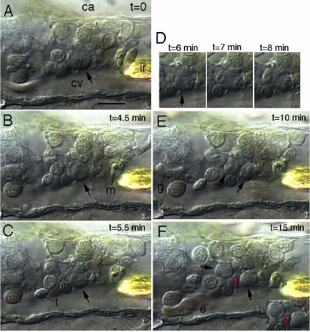
Fig. S1 Video-Enhanced Nomarski Images of the Mitosis of a Progenitor in the Caudal Hematopoietic Tissue at 4 dpf Around the cell undergoing mitosis (arrows) appear various other progenitors, plus a macrophage (m) and a granulocyte (g) which migrated away between E and F. (A) Prophase; (B) Metaphase; (C) Anaphase A; (D) Anaphase B and telophase; (E) Daughter cells with condensed chromatin and no nucleus yet; (F) Nuclear assembly almost complete; the two daughter cells are still connected by a tether (red arrow), better seen in the lower right inset; note the deformation of a circulating erythrocyte (e) as it bumped in a progenitor, demonstrating that the latter is intraluminal. Ir, iridophore; bar, 10 μm.
Reprinted from Immunity, 25(6), Murayama, E., Kissa, K., Zapata, A., Mordelet, E., Briolat, V., Lin, H.F., Handin, R.I., and Herbomel, P., Tracing Hematopoietic Precursor Migration to Successive Hematopoietic Organs during Zebrafish Development, 963-975, Copyright (2006) with permission from Elsevier. Full text @ Immunity