
IMAGE
Fig. 4
- ID
- ZDB-IMAGE-071223-20
- Genes
- Publication
- Nogare et al., 2007 - Zebrafish cdc25a is expressed during early development and limiting for post-blastoderm cell cycle progression
- All Figures
- Figures for Nogare et al., 2007
Image
|
Figure Caption
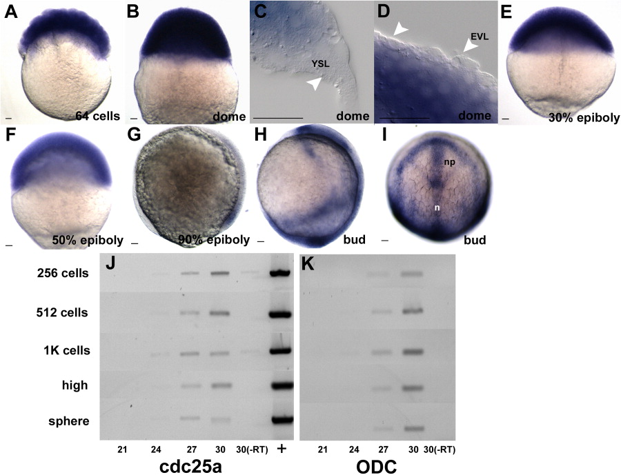
Fig. 4 In situ hybridization using a cdc25a antisense probe during epiboly. Represented are staged semi-quantitative RT-PCR results using either cdc25a (j) or ODC (k) specific primers. Cycle numbers are as indicated below panels, with stages at left. (+) plasmid positive control; (-RT) no RT control reactions. All embryos are oriented animal pole up. g and h are lateral views with the dorsal side to the right and i is a dorsal view. YSL, yolk syncytial layer; EVL, enveloping layer; n, notochord; np, neural plate. Scale bar = 20 μm.
Figure Data
Acknowledgments
This image is the copyrighted work of the attributed author or publisher, and
ZFIN has permission only to display this image to its users.
Additional permissions should be obtained from the applicable author or publisher of the image.
Full text @ Dev. Dyn.