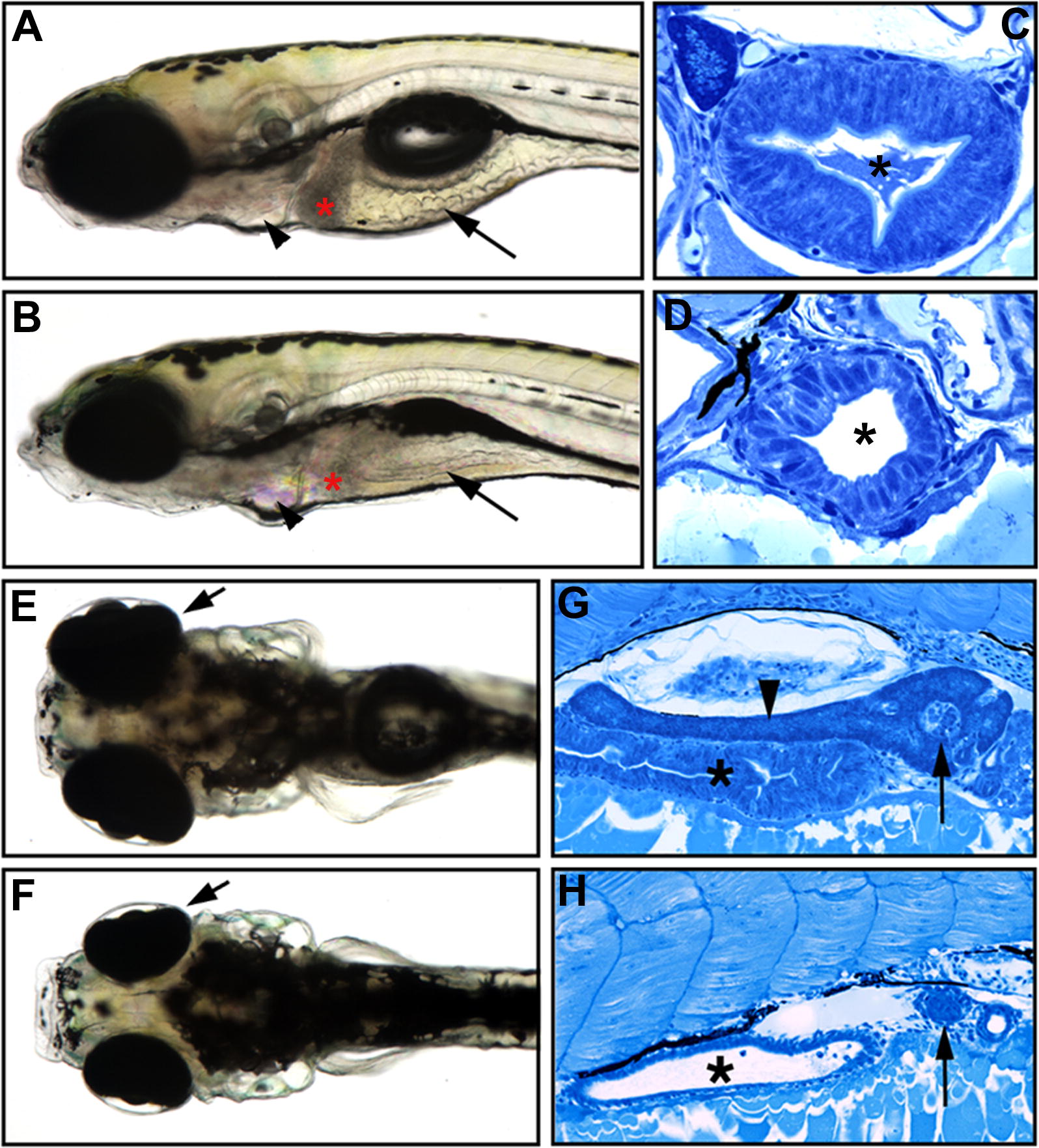
Fig. 1 The slj Mutation Disrupts Intestinal and Exocrine Pancreas Development (A–D) Lateral views of 5-dpf wild-type (A) and slj (B) larvae, with representative histological cross-sections (C and D). The slj intestine is small, thin walled, and lacks folds (arrows, [A and B]) compared with wild type, and the slj terminal branchial arches are reduced (arrowheads). The columnar morphology and apical microvilli of the slj epithelium are less developed than in wild-type larvae (C and D); an asterisk (*) indicates the intestinal lumen. (E and F) The size of the slj liver (indicated by red asterisk in [A] and [B]) and the retinae (arrows, [E and F], dorsal view) are also reduced. (G and H) Although exocrine tissue is well developed in wild-type embryos (arrowhead in [G]), it is markedly reduced or absent in slj embryos (H); asterisks indicate intestine. By contrast, the slj and wild-type pancreatic islets are of comparable size (arrows).
Image
Figure Caption
Figure Data
Acknowledgments
This image is the copyrighted work of the attributed author or publisher, and
ZFIN has permission only to display this image to its users.
Additional permissions should be obtained from the applicable author or publisher of the image.
Full text @ PLoS Biol.

