
IMAGE
Fig. 1
- ID
- ZDB-IMAGE-071115-12
- Publication
- Crotwell et al., 2007 - Gene expression patterns underlying proximal-distal skeletal segmentation in late-stage zebrafish, Danio rerio
- All Figures
- Figures for Crotwell et al., 2007
Image
|
Figure Caption
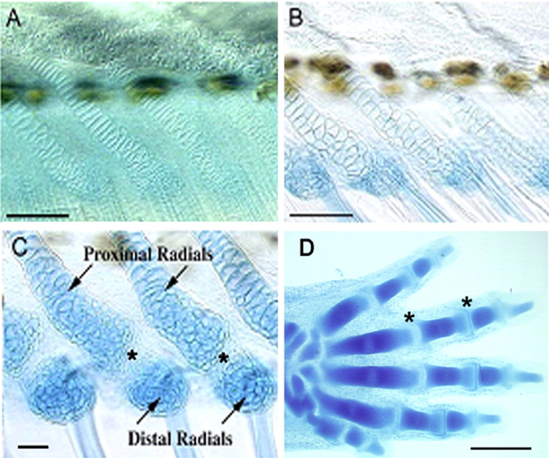
Fig. 1 A-D: Cleared and cartilage-stained skeletal preparations showing the progression of segmentation in zebrafish anal fin radials (A-C; adapted from Bird and Mabee,[2003]) and an 18 days postfertilization mouse embryo (D). A: 5.4 mm. B: 5.6 mm. C: 6.5 mm, proximal and distal radials are identified; asterisks mark the zones of segmentation. D: The mouse left forehand is shown here in a dorsal view with anterior at bottom. Two joints in digit four are marked with asterisks. Scale bars = 0.5 mm.
Acknowledgments
This image is the copyrighted work of the attributed author or publisher, and
ZFIN has permission only to display this image to its users.
Additional permissions should be obtained from the applicable author or publisher of the image.
Full text @ Dev. Dyn.