
Fig. 3
- ID
- ZDB-IMAGE-071112-29
- Genes
- Publication
- Ott et al., 2007 - Characterization of the enigma family in zebrafish
- All Figures
- Figures for Ott et al., 2007
|
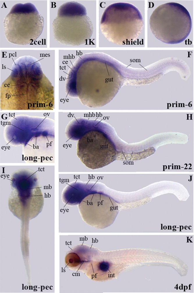
Fig. 3 A-K: Gene expression patterns of enigma variants encoding the LIM domain by whole-mount in situ hybridization. Similar to the PDZ domain encoding variant, eight different developmental stages are shown. We present embryos at the two-cell stage during the cleavage period until at 4 days postfertilization (dpf) at the early larval period. F,H,J,K: Lateral views are shown for embryos at prim-6, prim-22, long-pec, and 4 dpf stages. F,G: A higher magnification of the dorsal view at prim-6 is shown in F and of the lateral view at the long-pec stage in G. The long-pec stage is also shown in I from a dorsal view. ba, branchial arches; ce, cerebellum; cm, cephalic musculature; dv, diencephalic vein; fp, floor plate; hb, hindbrain; int, intestine; ls, lens; mes, mesencephalon; mb, midbrain; mhb, midbrain-hindbrain boundary; ov, otic vesicle; pf, pectoral fin buds; pcl, proliferative cell layer; som, somites; tct, tectum; tgm, tegmentum.